7JZ4

Crystal structure of broadly Plasmodium RIFIN reactive LAIR1-inserted antibody MGD21


Experimental Data Snapshot

  • Method: X-RAY DIFFRACTION
  • Resolution: 2.75 Å
  • R-Value Free: 0.249 
  • R-Value Work: 0.194 
  • R-Value Observed: 0.197 

wwPDB Validation   3D Report Full Report


Ligand Structure Quality Assessment 


This is version 1.2 of the entry. See complete history


Literature

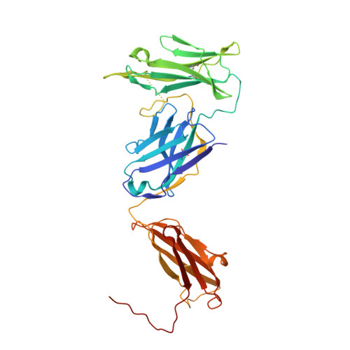
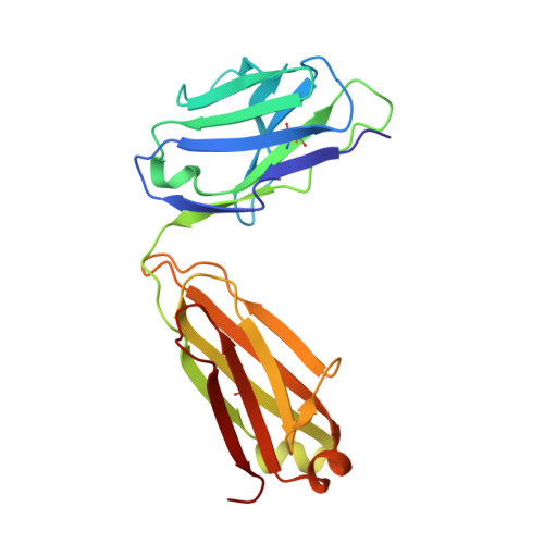
Structural basis of LAIR1 targeting by polymorphic Plasmodium RIFINs.

Xu, K.Wang, Y.Shen, C.H.Chen, Y.Zhang, B.Liu, K.Tsybovsky, Y.Wang, S.Farney, S.K.Gorman, J.Stephens, T.Verardi, R.Yang, Y.Zhou, T.Chuang, G.Y.Lanzavecchia, A.Piccoli, L.Kwong, P.D.

(2021) Nat Commun 12: 4226-4226

  • DOI: https://doi.org/10.1038/s41467-021-24291-6
  • Primary Citation of Related Structures:  
    7JZ1, 7JZ4, 7JZI, 7JZK

  • PubMed Abstract: 

    RIFIN, a large family of Plasmodium variant surface antigens, plays a crucial role in malaria pathogenesis by mediating immune suppression through activation of inhibitory receptors such as LAIR1, and antibodies with LAIR1 inserts have been identified that bind infected erythrocytes through RIFIN. However, details of RIFIN-mediated LAIR1 recognition and receptor activation have been unclear. Here, we use negative-stain EM to define the architecture of LAIR1-inserted antibodies and determine crystal structures of RIFIN-variable 2 (V2) domain in complex with a LAIR1 domain. These structures reveal the LAIR1-binding region of RIFIN to be hydrophobic and membrane-distal, to exhibit extensive structural diversity, and to interact with RIFIN-V2 in a one-to-one fashion. Through structural and sequence analysis of various LAIR1 constructs, we identify essential elements of RIFIN-binding on LAIR1. Furthermore, a structure-derived LAIR1-binding sequence signature ascertained >20 LAIR1-binding RIFINs, including some from P. falciparum field strains and Plasmodium species infecting gorillas and chimpanzees.


  • Organizational Affiliation

    Vaccine Research Center, National Institute of Allergy and Infectious Diseases, National Institutes of Health, Bethesda, MD, USA. xu.4692@osu.edu.


Macromolecules
Find similar proteins by:  (by identity cutoff)  |  3D Structure
Entity ID: 1
MoleculeChains Sequence LengthOrganismDetailsImage
MGD21 heavy chainA,
D [auth C]
364Homo sapiensMutation(s): 0 
Entity Groups  
Sequence Clusters30% Identity50% Identity70% Identity90% Identity95% Identity100% Identity
Sequence Annotations
Expand
  • Reference Sequence
Find similar proteins by:  (by identity cutoff)  |  3D Structure
Entity ID: 2
MoleculeChains Sequence LengthOrganismDetailsImage
MGD21 light chainB,
C [auth D]
213Homo sapiensMutation(s): 0 
Entity Groups  
Sequence Clusters30% Identity50% Identity70% Identity90% Identity95% Identity100% Identity
Sequence Annotations
Expand
  • Reference Sequence
Experimental Data & Validation

Experimental Data

  • Method: X-RAY DIFFRACTION
  • Resolution: 2.75 Å
  • R-Value Free: 0.249 
  • R-Value Work: 0.194 
  • R-Value Observed: 0.197 
  • Space Group: P 1 21 1
Unit Cell:
Length ( Å )Angle ( ˚ )
a = 80.932α = 90
b = 81.099β = 94.03
c = 111.561γ = 90
Software Package:
Software NamePurpose
PHENIXrefinement
SCALEPACKdata scaling
PDB_EXTRACTdata extraction
HKL-2000data reduction
PHASERphasing

Structure Validation

View Full Validation Report


Ligand Structure Quality Assessment 


Entry History 

Deposition Data

Revision History  (Full details and data files)

  • Version 1.0: 2021-05-26
    Type: Initial release
  • Version 1.1: 2021-09-22
    Changes: Database references
  • Version 1.2: 2023-10-18
    Changes: Data collection, Refinement description