7FF7 | pdb_00007ff7

Structure of OmpF2


Experimental Data Snapshot

  • Method: X-RAY DIFFRACTION
  • Resolution: 3.38 Å
  • R-Value Free: 
    0.337 (Depositor), 0.326 (DCC) 
  • R-Value Work: 
    0.290 (Depositor), 0.285 (DCC) 
  • R-Value Observed: 
    0.292 (Depositor) 

Starting Model: experimental
View more details

wwPDB Validation 3D Report Full Report

Validation slider image for 7FF7

This is version 1.2 of the entry. See complete history

Literature

Design and directed evolution of noncanonical beta-stereoselective metalloglycosidases.

Jeong, W.J.Song, W.J.

(2022) Nat Commun 13: 6844-6844

  • DOI: https://doi.org/10.1038/s41467-022-34713-8
  • Primary Citation Related Structures: 
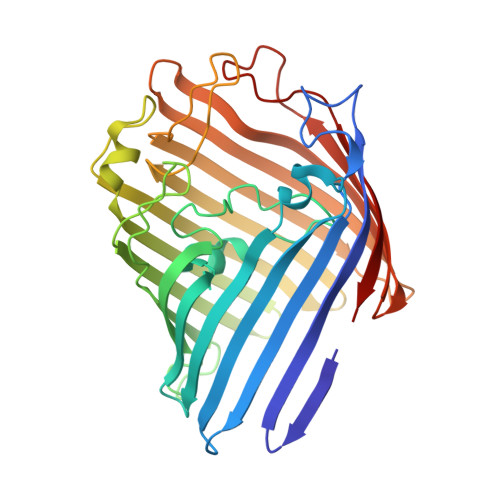
    7FDY, 7FF7

  • PubMed Abstract: 

    Metallohydrolases are ubiquitous in nearly all subclasses of hydrolases, utilizing metal elements to activate a water molecule and facilitate its subsequent dissociation of diverse chemical bonds. However, such a catalytic role of metal ions is rarely found with glycosidases that hydrolyze the glycosidic bonds in sugars. Herein, we design metalloglycosidases by constructing a hydrolytically active Zn-binding site within a barrel-shaped outer membrane protein OmpF. Structure- and mechanism-based redesign and directed evolution have led to the emergence of Zn-dependent glycosidases with catalytic proficiency of 2.8 × 10 9 and high β-stereoselectivity. Biochemical characterizations suggest that the Zn-binding site constitutes a key catalytic motif along with at least one adjacent acidic residue. This work demonstrates that unprecedented metalloenzymes can be tailor-made, expanding the scope of inorganic reactivities in proteinaceous environments, resetting the structural and functional diversity of metalloenzymes, and providing the potential molecular basis of unidentified metallohydrolases and novel whole-cell biocatalysts.


  • Organizational Affiliation
    • Department of Chemistry, Seoul National University, Seoul, 08826, Republic of Korea.

Macromolecule Content 

  • Total Structure Weight: 111.48 kDa 
  • Atom Count: 7,055 
  • Modeled Residue Count: 986 
  • Deposited Residue Count: 1,020 
  • Unique protein chains: 1

Macromolecules

Find similar proteins by:|  3D Structure
Entity ID: 1
MoleculeChains  Sequence LengthOrganismDetailsImage
Outer membrane protein F
A, B, C
340Escherichia coliMutation(s): 0 
Gene Names: ompF_1SAMEA3751407_00438

Experimental Data & Validation

Experimental Data

  • Method: X-RAY DIFFRACTION
  • Resolution: 3.38 Å
  • R-Value Free:  0.337 (Depositor), 0.326 (DCC) 
  • R-Value Work:  0.290 (Depositor), 0.285 (DCC) 
  • R-Value Observed: 0.292 (Depositor) 
Space Group: C 1 2 1
Unit Cell:
Length ( Å )Angle ( ˚ )
a = 153.732α = 90
b = 111.968β = 110.87
c = 110.873γ = 90
Software Package:
Software NamePurpose
REFMACrefinement
HKL-2000data scaling
PDB_EXTRACTdata extraction
HKL-2000data reduction
MOLREPphasing

Structure Validation

View Full Validation Report


Entry History 

& Funding Information

Deposition Data


Funding OrganizationLocationGrant Number
Not fundedKorea, Republic Of2019R1C1C1003863

Revision History  (Full details and data files)

  • Version 1.0: 2022-11-16
    Type: Initial release
  • Version 1.1: 2023-04-19
    Changes: Database references
  • Version 1.2: 2023-11-29
    Changes: Data collection, Refinement description