7EN3 | pdb_00007en3

Crystal structure of tubulin in complex with Tubulysin analogue TGL


Experimental Data Snapshot

  • Method: X-RAY DIFFRACTION
  • Resolution: 2.64 Å
  • R-Value Free: 
    0.239 (Depositor), 0.227 (DCC) 
  • R-Value Work: 
    0.199 (Depositor) 
  • R-Value Observed: 
    0.201 (Depositor) 

Starting Model: experimental
View more details

wwPDB Validation 3D Report Full Report

Validation slider image for 7EN3

Ligand Structure Quality Assessment 


This is version 1.2 of the entry. See complete history

Literature

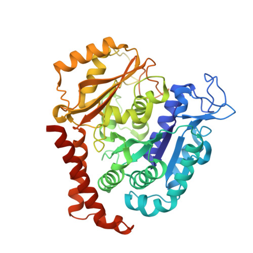
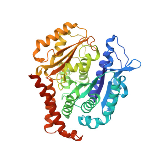
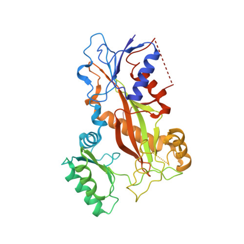
The X-ray structure of tubulysin analogue TGL in complex with tubulin and three possible routes for the development of next-generation tubulysin analogues.

Li, W.Tan, L.Zhang, Z.Xia, Q.Lei, D.Li, Y.Zhang, T.Zeng, S.Sima, X.Wang, Y.

(2021) Biochem Biophys Res Commun 565: 29-35

  • DOI: https://doi.org/10.1016/j.bbrc.2021.05.086
  • Primary Citation Related Structures: 
    7EN3

  • PubMed Abstract: 

    Microtubule-targeting agents (MTAs) are the most commonly used anti-cancer drugs. At least fourteen microtubule inhibitors and ten antibody drug conjugates (ADCs) linking MTAs are approved by FDA for clinical use in cancer therapy. In current research, we determined the crystal structure of tubulysin analogue TGL in complex with tubulin at a high resolution (2.65 Å). In addition, we summarized all of the previously published high-resolution crystal structures of ligands in the vinca site to provide structural insights for the rational design of the new vinca-site ligands. Moreover, based on the aligned results of the vinca site ligands, we provided three possible routes for designing new tubulysin analogues, namely macrocyclization between the N-14 side chain and the N-9 side chain, the hybird of tubulysin M and phomopsin A, and growing new aryl group at C-21. These designed structures will inspire the development of new MTAs or payloads in cancer therapy.


  • Organizational Affiliation
    • State Key Laboratory of Biotherapy and Collaborative Innovation Center for Biotherapy, West China Hospital, Sichuan University, Chengdu, 610041, People's Republic of China. Electronic address: liwentingo@qq.com.

Macromolecule Content 

  • Total Structure Weight: 265.62 kDa 
  • Atom Count: 17,798 
  • Modeled Residue Count: 2,188 
  • Deposited Residue Count: 2,312 
  • Unique protein chains: 4

Macromolecules

Find similar proteins by:|  3D Structure
Entity ID: 1
MoleculeChains  Sequence LengthOrganismDetailsImage
Tubulin alpha-1B chain
A, C
451Sus scrofaMutation(s): 0 
Gene Names: TUBA1B
EC: 3.6.5
UniProt
Find proteins for Q2XVP4 (Sus scrofa)
Explore Q2XVP4 
Go to UniProtKB:  Q2XVP4
Entity Groups
Sequence Clusters30% Identity50% Identity70% Identity90% Identity95% Identity100% Identity
UniProt GroupQ2XVP4
Sequence Annotations
Expand
Reference Sequence
Find similar proteins by:|  3D Structure
Entity ID: 2
MoleculeChains  Sequence LengthOrganismDetailsImage
Tubulin beta-2B chain
B, D
445Bos taurusMutation(s): 0 
Gene Names: TUBB2B
UniProt
Find proteins for Q6B856 (Bos taurus)
Explore Q6B856 
Go to UniProtKB:  Q6B856
Entity Groups
Sequence Clusters30% Identity50% Identity70% Identity90% Identity95% Identity100% Identity
UniProt GroupQ6B856
Sequence Annotations
Expand
Reference Sequence
Find similar proteins by:|  3D Structure
Entity ID: 3
MoleculeChains  Sequence LengthOrganismDetailsImage
Stathmin-4136Rattus norvegicusMutation(s): 0 
Gene Names: Stmn4
UniProt
Find proteins for P63043 (Rattus norvegicus)
Explore P63043 
Go to UniProtKB:  P63043
Entity Groups
Sequence Clusters30% Identity50% Identity70% Identity90% Identity95% Identity100% Identity
UniProt GroupP63043
Sequence Annotations
Expand
Reference Sequence
Find similar proteins by:|  3D Structure
Entity ID: 4
MoleculeChains  Sequence LengthOrganismDetailsImage
tubulin tyrosine ligase384Gallus gallusMutation(s): 0 
Gene Names: TTL

Small Molecules

Ligands 7 Unique
IDChains Name / Formula / InChI Key2D Diagram3D Interactions
J6R
(Subject of Investigation/LOI)

Query on J6R


Download:Ideal Coordinates CCD File
N [auth B],
T [auth D]
(2S,4R)-5-(4-fluorophenyl)-2-methyl-4-[[2-[(1R,3R)-4-methyl-3-[5-methylhexyl-[(2S,3S)-3-methyl-2-[[(2R)-1-methylpiperidin-2-yl]carbonylamino]pentanoyl]amino]-1-oxidanyl-pentyl]-1,3-thiazol-4-yl]carbonylamino]pentanoic acid
C42 H66 F N5 O6 S
XFQABYNPLHNBRH-KHUAJKBRSA-N
GTP

Query on GTP


Download:Ideal Coordinates CCD File
G [auth A],
P [auth C]
GUANOSINE-5'-TRIPHOSPHATE
C10 H16 N5 O14 P3
XKMLYUALXHKNFT-UUOKFMHZSA-N
ACP

Query on ACP


Download:Ideal Coordinates CCD File
U [auth F]PHOSPHOMETHYLPHOSPHONIC ACID ADENYLATE ESTER
C11 H18 N5 O12 P3
UFZTZBNSLXELAL-IOSLPCCCSA-N
GDP

Query on GDP


Download:Ideal Coordinates CCD File
J [auth B],
S [auth D]
GUANOSINE-5'-DIPHOSPHATE
C10 H15 N5 O11 P2
QGWNDRXFNXRZMB-UUOKFMHZSA-N
MES

Query on MES


Download:Ideal Coordinates CCD File
L [auth B],
M [auth B],
O [auth C]
2-(N-MORPHOLINO)-ETHANESULFONIC ACID
C6 H13 N O4 S
SXGZJKUKBWWHRA-UHFFFAOYSA-N
CA

Query on CA


Download:Ideal Coordinates CCD File
I [auth A],
R [auth C]
CALCIUM ION
Ca
BHPQYMZQTOCNFJ-UHFFFAOYSA-N
MG

Query on MG


Download:Ideal Coordinates CCD File
H [auth A],
K [auth B],
Q [auth C]
MAGNESIUM ION
Mg
JLVVSXFLKOJNIY-UHFFFAOYSA-N

Experimental Data & Validation

Experimental Data

  • Method: X-RAY DIFFRACTION
  • Resolution: 2.64 Å
  • R-Value Free:  0.239 (Depositor), 0.227 (DCC) 
  • R-Value Work:  0.199 (Depositor) 
  • R-Value Observed: 0.201 (Depositor) 
Space Group: P 21 21 21
Unit Cell:
Length ( Å )Angle ( ˚ )
a = 104.607α = 90
b = 154.216β = 90
c = 186.865γ = 90
Software Package:
Software NamePurpose
REFMACrefinement
HKL-2000data reduction
PDB_EXTRACTdata extraction
HKL-2000data scaling
MOLREPphasing

Structure Validation

View Full Validation Report


Ligand Structure Quality Assessment 


Entry History 

& Funding Information

Deposition Data


Funding OrganizationLocationGrant Number
National Natural Science Foundation of China (NSFC)China81703553

Revision History  (Full details and data files)

  • Version 1.0: 2022-04-27
    Type: Initial release
  • Version 1.1: 2022-05-04
    Changes: Database references
  • Version 1.2: 2023-11-29
    Changes: Data collection, Refinement description