


Structural analysis of EcoT38I restriction endonuclease
Kita, K., Mikami, B.To be published.
Experimental Data Snapshot
Starting Model: experimental
View more details
Entity ID: 1 | |||||
|---|---|---|---|---|---|
| Molecule | Chains | Sequence Length | Organism | Details | Image |
| EcoT38I restriction endonuclease | 351 | Peduovirus P2 | Mutation(s): 0 Gene Names: ecoT38IR | 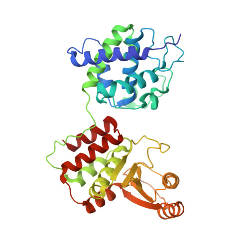 | |
UniProt | |||||
Entity Groups | |||||
| Sequence Clusters | 30% Identity50% Identity70% Identity90% Identity95% Identity100% Identity | ||||
| UniProt Group | Q83VS8 | ||||
Sequence AnnotationsExpand | |||||
Reference Sequence | |||||
Entity ID: 2 | ||||
| Molecule | Chains | Length | Organism | Image |
|---|---|---|---|---|
| DNA (5'-D(*GP*GP*CP*AP*GP*AP*GP*CP*TP*CP*AP*CP*G)-3') | C [auth E] | 13 | Escherichia coli | 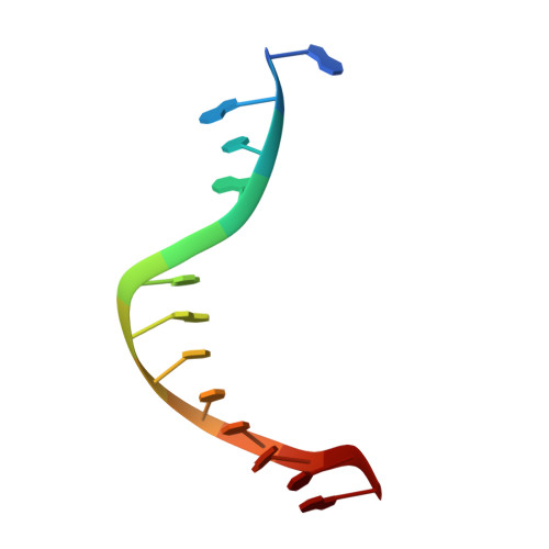 |
Sequence AnnotationsExpand | ||||
Reference Sequence | ||||
Entity ID: 3 | ||||
| Molecule | Chains | Length | Organism | Image |
|---|---|---|---|---|
| DNA (5'-D(P*CP*GP*TP*GP*AP*GP*CP*TP*CP*TP*GP*C)-3') | D [auth F] | 13 | Escherichia coli | 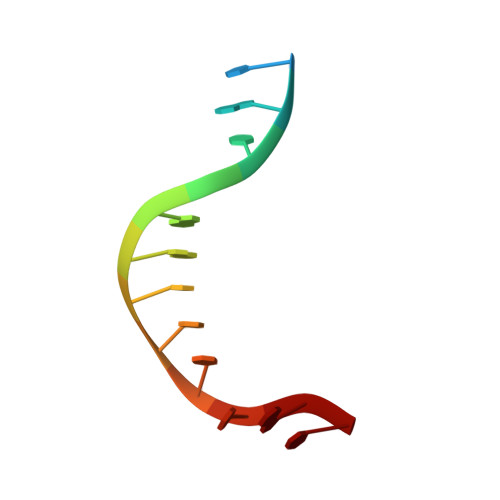 |
Sequence AnnotationsExpand | ||||
Reference Sequence | ||||
| Ligands 6 Unique | |||||
|---|---|---|---|---|---|
| ID | Chains | Name / Formula / InChI Key | 2D Diagram | 3D Interactions | |
| PGE (Subject of Investigation/LOI) Download:Ideal Coordinates CCD File | P [auth B] | TRIETHYLENE GLYCOL C6 H14 O4 ZIBGPFATKBEMQZ-UHFFFAOYSA-N |  | ||
| TRS (Subject of Investigation/LOI) Download:Ideal Coordinates CCD File | G [auth A], H [auth A], Q [auth B], R [auth B] | 2-AMINO-2-HYDROXYMETHYL-PROPANE-1,3-DIOL C4 H12 N O3 LENZDBCJOHFCAS-UHFFFAOYSA-O |  | ||
| GOL (Subject of Investigation/LOI) Download:Ideal Coordinates CCD File | I [auth A] J [auth A] K [auth A] L [auth A] S [auth B] | GLYCEROL C3 H8 O3 PEDCQBHIVMGVHV-UHFFFAOYSA-N |  | ||
| CA (Subject of Investigation/LOI) Download:Ideal Coordinates CCD File | E [auth A], F [auth A], N [auth B], W [auth F] | CALCIUM ION Ca BHPQYMZQTOCNFJ-UHFFFAOYSA-N |  | ||
| CL (Subject of Investigation/LOI) Download:Ideal Coordinates CCD File | M [auth A], X [auth F] | CHLORIDE ION Cl VEXZGXHMUGYJMC-UHFFFAOYSA-M |  | ||
| NA (Subject of Investigation/LOI) Download:Ideal Coordinates CCD File | O [auth B] | SODIUM ION Na FKNQFGJONOIPTF-UHFFFAOYSA-N |  | ||
| Length ( Å ) | Angle ( ˚ ) |
|---|---|
| a = 156.291 | α = 90 |
| b = 156.291 | β = 90 |
| c = 173.049 | γ = 120 |
| Software Name | Purpose |
|---|---|
| PHENIX | refinement |
| XDS | data reduction |
| XDS | data scaling |
| MOLREP | phasing |
| Funding Organization | Location | Grant Number |
|---|---|---|
| Japan Society for the Promotion of Science (JSPS) | Japan | 16380061 |


