7CU5 | pdb_00007cu5

N-Glycosylation of PD-1 and glycosylation dependent binding of PD-1 specific monoclonal antibody camrelizumab


Experimental Data Snapshot

  • Method: X-RAY DIFFRACTION
  • Resolution: 2.81 Å
  • R-Value Free: 
    0.252 (Depositor), 0.253 (DCC) 
  • R-Value Work: 
    0.212 (Depositor), 0.214 (DCC) 
  • R-Value Observed: 
    0.214 (Depositor) 

Starting Model: experimental
View more details

wwPDB Validation 3D Report Full Report

Validation slider image for 7CU5

Ligand Structure Quality Assessment 


This is version 1.3 of the entry. See complete history

Literature

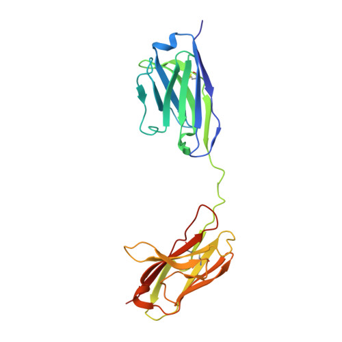
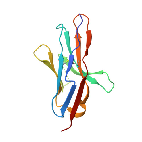
N-glycosylation of PD-1 promotes binding of camrelizumab.

Liu, K.Tan, S.Jin, W.Guan, J.Wang, Q.Sun, H.Qi, J.Yan, J.Chai, Y.Wang, Z.Deng, C.Gao, G.F.

(2020) EMBO Rep 21: e51444-e51444

  • DOI: https://doi.org/10.15252/embr.202051444
  • Primary Citation Related Structures: 
    7CU5

  • PubMed Abstract: 

    PD-1 is a highly glycosylated inhibitory receptor expressed mainly on T cells. Targeting of PD-1 with monoclonal antibodies (MAbs) to block the interaction with its ligand PD-L1 has been successful for the treatment of multiple tumors. However, polymorphisms at N-glycosylation sites of PD-1 exist in the human population that might affect antibody binding, and dysregulated glycosylation has been observed in the tumor microenvironment. Here, we demonstrate varied N-glycan composition in PD-1, and show that the binding affinity of camrelizumab, a recently approved PD-1-specific MAb, to non-glycosylated PD-1 proteins from E. coli is substantially decreased compared with glycosylated PD-1. The structure of the camrelizumab/PD-1 complex reveals that camrelizumab mainly utilizes its heavy chain to bind to PD-1, while the light chain sterically inhibits the binding of PD-L1 to PD-1. Glycosylation of asparagine 58 (N58) promotes the interaction with camrelizumab, while the efficiency of camrelizumab to inhibit the binding of PD-L1 is substantially reduced for glycosylation-deficient PD-1. These results increase our understanding of how glycosylation affects the activity of PD-1-specific MAbs during immune checkpoint therapy.


  • Organizational Affiliation
    • Faculty of Health Sciences, University of Macau, Macau SAR, China.

Macromolecule Content 

  • Total Structure Weight: 79.83 kDa 
  • Atom Count: 5,486 
  • Modeled Residue Count: 682 
  • Deposited Residue Count: 696 
  • Unique protein chains: 2

Macromolecules

Find similar proteins by:|  3D Structure
Entity ID: 1
MoleculeChains  Sequence LengthOrganismDetailsImage
camrelizumab-scFvA [auth B],
C [auth A]
230Homo sapiensMutation(s): 0 
Find similar proteins by:|  3D Structure
Entity ID: 2
MoleculeChains  Sequence LengthOrganismDetailsImage
Programmed cell death protein 1B [auth E],
D [auth Q]
118Homo sapiensMutation(s): 0 
Gene Names: PDCD1PD1
UniProt & NIH Common Fund Data Resources
Find proteins for Q15116 (Homo sapiens)
Explore Q15116 
Go to UniProtKB:  Q15116
PHAROS:  Q15116
GTEx:  ENSG00000188389 
Entity Groups
Sequence Clusters30% Identity50% Identity70% Identity90% Identity95% Identity100% Identity
UniProt GroupQ15116
Glycosylation
Glycosylation Sites: 4Go to GlyGen: Q15116-1
Sequence Annotations
Expand
Reference Sequence

Oligosaccharides

Help  
Entity ID: 3
MoleculeChains Length2D Diagram GlycosylationD Interactions
alpha-D-mannopyranose-(1-6)-alpha-D-mannopyranose-(1-4)-2-acetamido-2-deoxy-beta-D-glucopyranose-(1-4)-[alpha-L-fucopyranose-(1-6)]2-acetamido-2-deoxy-beta-D-glucopyranoseE [auth D]5N-Glycosylation
Glycosylation Resources
GlyTouCan: G17392HI
GlyCosmos: G17392HI
GlyGen: G17392HI
Entity ID: 4
MoleculeChains Length2D Diagram GlycosylationD Interactions
2-acetamido-2-deoxy-beta-D-glucopyranose-(1-4)-[alpha-L-fucopyranose-(1-6)]2-acetamido-2-deoxy-beta-D-glucopyranoseF [auth K],
H [auth I]
3N-Glycosylation
Glycosylation Resources
GlyTouCan: G21290RB
GlyCosmos: G21290RB
GlyGen: G21290RB
Entity ID: 5
MoleculeChains Length2D Diagram GlycosylationD Interactions
alpha-D-mannopyranose-(1-3)-alpha-D-mannopyranose-(1-4)-2-acetamido-2-deoxy-beta-D-glucopyranose-(1-4)-[alpha-L-fucopyranose-(1-6)]2-acetamido-2-deoxy-beta-D-glucopyranoseG [auth C]5N-Glycosylation
Glycosylation Resources
GlyTouCan: G26032DN
GlyCosmos: G26032DN
GlyGen: G26032DN

Experimental Data & Validation

Experimental Data

  • Method: X-RAY DIFFRACTION
  • Resolution: 2.81 Å
  • R-Value Free:  0.252 (Depositor), 0.253 (DCC) 
  • R-Value Work:  0.212 (Depositor), 0.214 (DCC) 
  • R-Value Observed: 0.214 (Depositor) 
Space Group: P 1
Unit Cell:
Length ( Å )Angle ( ˚ )
a = 43.108α = 69.381
b = 77.622β = 78.444
c = 96.051γ = 88.566
Software Package:
Software NamePurpose
PHENIXrefinement
HKL-2000data reduction
HKL-2000data scaling
PHASERphasing

Structure Validation

View Full Validation Report


Ligand Structure Quality Assessment 


Entry History 

Deposition Data

Revision History  (Full details and data files)

  • Version 1.0: 2020-10-14
    Type: Initial release
  • Version 1.1: 2021-03-31
    Changes: Database references
  • Version 1.2: 2023-11-29
    Changes: Data collection, Database references, Refinement description
  • Version 1.3: 2024-10-30
    Changes: Structure summary