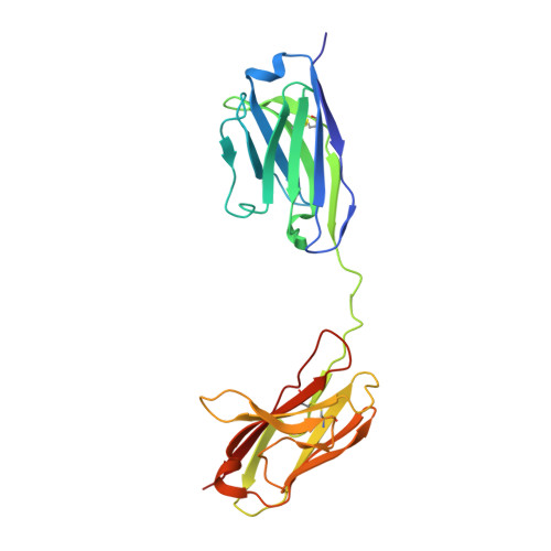
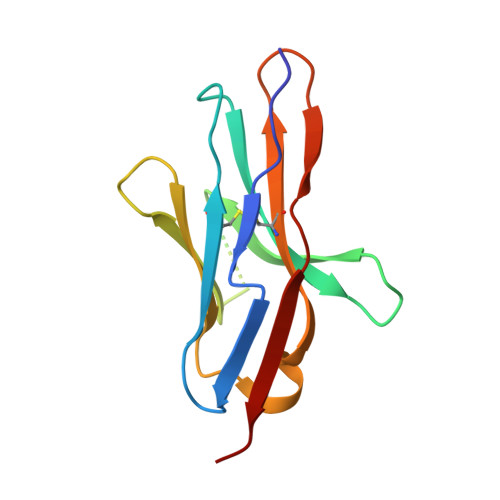
7CU5 | pdb_00007cu5

N-Glycosylation of PD-1 and glycosylation dependent binding of PD-1 specific monoclonal antibody camrelizumab


Experimental Data Snapshot

  • Method: X-RAY DIFFRACTION
  • Resolution: 2.81 Å
  • R-Value Free: 
    0.252 (Depositor), 0.253 (DCC) 
  • R-Value Work: 
    0.212 (Depositor), 0.214 (DCC) 
  • R-Value Observed: 
    0.214 (Depositor) 

Starting Model: experimental
View more details

wwPDB Validation 3D Report Full Report

Validation slider image for 7CU5

Ligand Structure Quality Assessment 


This is version 1.3 of the entry. See complete history. 

Literature

N-glycosylation of PD-1 promotes binding of camrelizumab.

Liu, K., Tan, S., Jin, W., Guan, J., Wang, Q., Sun, H., Qi, J., Yan, J., Chai, Y., Wang, Z., Deng, C., Gao, G.F.

(2020) EMBO Rep 21: e51444-e51444

  • DOI: https://doi.org/10.15252/embr.202051444
  • Primary Citation Related Structures: 
    7CU5

  • PubMed Abstract: 

    PD-1 is a highly glycosylated inhibitory receptor expressed mainly on T cells. Targeting of PD-1 with monoclonal antibodies (MAbs) to block the interaction with its ligand PD-L1 has been successful for the treatment of multiple tumors. However, polymorphisms at N-glycosylation sites of PD-1 exist in the human population that might affect antibody binding, and dysregulated glycosylation has been observed in the tumor microenvironment. Here, we demonstrate varied N-glycan composition in PD-1, and show that the binding affinity of camrelizumab, a recently approved PD-1-specific MAb, to non-glycosylated PD-1 proteins from E. coli is substantially decreased compared with glycosylated PD-1. The structure of the camrelizumab/PD-1 complex reveals that camrelizumab mainly utilizes its heavy chain to bind to PD-1, while the light chain sterically inhibits the binding of PD-L1 to PD-1. Glycosylation of asparagine 58 (N58) promotes the interaction with camrelizumab, while the efficiency of camrelizumab to inhibit the binding of PD-L1 is substantially reduced for glycosylation-deficient PD-1. These results increase our understanding of how glycosylation affects the activity of PD-1-specific MAbs during immune checkpoint therapy.


  • Organizational Affiliation: 
    • Faculty of Health Sciences, University of Macau, Macau SAR, China.

Macromolecule Content 

  • Total Structure Weight: 79.83 kDa 
  • Atom Count: 5,486 
  • Modeled Residue Count: 682 
  • Deposited Residue Count: 696 
  • Unique protein chains: 2

Macromolecules

Find similar proteins by:|  3D Structure
Entity ID: 1
MoleculeChains  Sequence LengthOrganismDetailsImage
camrelizumab-scFvA [auth B],
C [auth A]
230Homo sapiensMutation(s): 0 
Entity Groups
Sequence Clusters30% Identity50% Identity70% Identity90% Identity95% Identity100% Identity
Sequence Annotations
Expand
Reference Sequence
Find similar proteins by:|  3D Structure
Entity ID: 2
MoleculeChains  Sequence LengthOrganismDetailsImage
Programmed cell death protein 1B [auth E],
D [auth Q]
118Homo sapiensMutation(s): 0 
Gene Names: PDCD1, PD1
UniProt & NIH Common Fund Data Resources
Find proteins for Q15116 (Homo sapiens)
Explore Q15116 
Go to UniProtKB:  Q15116
PHAROS:  Q15116
GTEx:  ENSG00000188389 
Entity Groups
Sequence Clusters30% Identity50% Identity70% Identity90% Identity95% Identity100% Identity
UniProt GroupQ15116
Glycosylation
Glycosylation Sites: 4Go to GlyGen: Q15116-1
Sequence Annotations
Expand
Reference Sequence

Oligosaccharides

Help  
Entity ID: 3
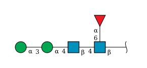
MoleculeChains Length2D Diagram GlycosylationD Interactions
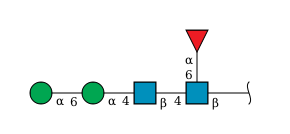
alpha-D-mannopyranose-(1-6)-alpha-D-mannopyranose-(1-4)-2-acetamido-2-deoxy-beta-D-glucopyranose-(1-4)-[alpha-L-fucopyranose-(1-6)]2-acetamido-2-deoxy-beta-D-glucopyranoseE [auth D]5N-Glycosylation
Glycosylation Resources
GlyTouCan: G17392HI
GlyCosmos: G17392HI
GlyGen: G17392HI
Entity ID: 4
MoleculeChains Length2D Diagram GlycosylationD Interactions
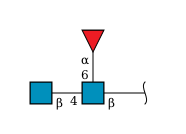
2-acetamido-2-deoxy-beta-D-glucopyranose-(1-4)-[alpha-L-fucopyranose-(1-6)]2-acetamido-2-deoxy-beta-D-glucopyranoseF [auth K],
H [auth I]
3N-Glycosylation
Glycosylation Resources
GlyTouCan: G21290RB
GlyCosmos: G21290RB
GlyGen: G21290RB
Entity ID: 5
MoleculeChains Length2D Diagram GlycosylationD Interactions
alpha-D-mannopyranose-(1-3)-alpha-D-mannopyranose-(1-4)-2-acetamido-2-deoxy-beta-D-glucopyranose-(1-4)-[alpha-L-fucopyranose-(1-6)]2-acetamido-2-deoxy-beta-D-glucopyranoseG [auth C]5N-Glycosylation
Glycosylation Resources
GlyTouCan: G26032DN
GlyCosmos: G26032DN
GlyGen: G26032DN

Experimental Data & Validation

Experimental Data

  • Method: X-RAY DIFFRACTION
  • Resolution: 2.81 Å
  • R-Value Free:  0.252 (Depositor), 0.253 (DCC) 
  • R-Value Work:  0.212 (Depositor), 0.214 (DCC) 
  • R-Value Observed: 0.214 (Depositor) 
Space Group: P 1
Unit Cell:
Length ( Å )Angle ( ˚ )
a = 43.108α = 69.381
b = 77.622β = 78.444
c = 96.051γ = 88.566
Software Package:
Software NamePurpose
PHENIXrefinement
HKL-2000data reduction
HKL-2000data scaling
PHASERphasing

Structure Validation

View Full Validation Report


Ligand Structure Quality Assessment 


Entry History 

Deposition Data

Revision History  (Full details and data files)

  • Version 1.0: 2020-10-14
    Type: Initial release
  • Version 1.1: 2021-03-31
    Changes: Database references
  • Version 1.2: 2023-11-29
    Changes: Data collection, Database references, Refinement description
  • Version 1.3: 2024-10-30
    Changes: Structure summary