7C6Z | pdb_00007c6z

Crystal structure of beta-glycosides-binding protein (W67A) of ABC transporter in an open state


Experimental Data Snapshot

  • Method: X-RAY DIFFRACTION
  • Resolution: 1.63 Å
  • R-Value Free: 
    0.196 (Depositor), 0.220 (DCC) 
  • R-Value Work: 
    0.158 (Depositor) 
  • R-Value Observed: 
    0.160 (Depositor) 

Starting Model: experimental
View more details

wwPDB Validation 3D Report Full Report

Validation slider image for 7C6Z

This is version 1.3 of the entry. See complete history

Literature

Conformational Trapping of a beta-Glucosides-Binding Protein Unveils the Selective Two-Step Ligand-Binding Mechanism of ABC Importers.

Chandravanshi, M.Samanta, R.Kanaujia, S.P.

(2020) J Mol Biology 432: 5711-5734

  • DOI: https://doi.org/10.1016/j.jmb.2020.08.021
  • Primary Citation Related Structures: 
    7C63, 7C64, 7C66, 7C67, 7C68, 7C69, 7C6F, 7C6G, 7C6H, 7C6I, 7C6J, 7C6K, 7C6L, 7C6M, 7C6N, 7C6R, 7C6T, 7C6V, 7C6W, 7C6X, 7C6Y, 7C6Z, 7C70, 7C71

  • PubMed Abstract: 

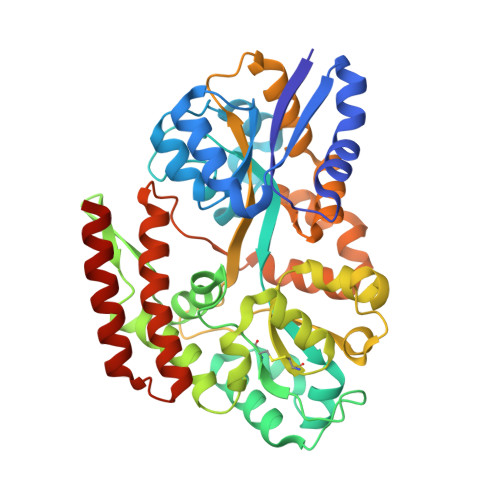
    Substrate-binding proteins (SBPs), selectively capture ligand(s) and ensure their translocation via its cognate ATP-binding cassette (ABC) import system. SBPs bind their cognate ligand(s) via an induced-fit mechanism known as the "Venus Fly-trap"; however, this mechanism lacks the atomic details of all conformational landscape as the confirmatory evidence(s) in its support. In this study, we delineate the atomic details of an SBP, β-glucosides-binding protein (βGlyBP) from Thermus thermophilus HB8. The protein βGlyBP is multi-specific and binds to different types of β-glucosides varying in their glycosidic linkages viz. β-1,2; β-1,3; β-1,4 and β-1,6 with a degree of polymerization of 2-5 glucosyl units. Structurally, the protein βGlyBP possesses four subdomains (N1, N2, C1 and C2). The unliganded protein βGlyBP remains in an open state, which closes upon binding to sophorose (SOP2), laminari-oligosaccharides (LAMn), cello-oligosaccharides (CELn), and gentiobiose (GEN2). This study reports, for the first time, four different structural states (open-unliganded, partial-open-unliganded, open-liganded and closed-liganded) of the protein βGlyBP, revealing its conformational changes upon ligand binding and suggesting a two-step induced-fit mechanism. Further, results suggest that the conformational changes of N1 and C1 subdomains drive the ligand binding, unlike that of the whole N- and C-terminal domains (NTD and CTD) as known in the "Venus Fly-trap" mechanism. Additionally, profiling of stereo-selection mechanism for α- and β-glucosides reveals that in the ligand-binding site four secondary structural elements (L1, H1, H2 and H3) drive the ligand selection. In summary, results demonstrate that the details of conformational changes and ligand selection are pre-encoded in the SBPs.


  • Organizational Affiliation
    • Department of Biosciences and Bioengineering, Indian Institute of Technology Guwahati, Guwahati 781039, Assam, India.

Macromolecule Content 

  • Total Structure Weight: 92.99 kDa 
  • Atom Count: 7,239 
  • Modeled Residue Count: 830 
  • Deposited Residue Count: 846 
  • Unique protein chains: 1

Macromolecules

Find similar proteins by:|  3D Structure
Entity ID: 1
MoleculeChains  Sequence LengthOrganismDetailsImage
Sugar ABC transporter, periplasmic sugar-binding protein
A, B
423Thermus thermophilus HB8Mutation(s): 1 
Gene Names: TTHB082
UniProt
Find proteins for Q53W80 (Thermus thermophilus (strain ATCC 27634 / DSM 579 / HB8))
Explore Q53W80 
Go to UniProtKB:  Q53W80
Entity Groups
Sequence Clusters30% Identity50% Identity70% Identity90% Identity95% Identity100% Identity
UniProt GroupQ53W80
Sequence Annotations
Expand
Reference Sequence

Small Molecules

Ligands 8 Unique
IDChains Name / Formula / InChI Key2D Diagram3D Interactions
PGE

Query on PGE


Download:Ideal Coordinates CCD File
K [auth A]TRIETHYLENE GLYCOL
C6 H14 O4
ZIBGPFATKBEMQZ-UHFFFAOYSA-N
PEG

Query on PEG


Download:Ideal Coordinates CCD File
J [auth A],
S [auth B]
DI(HYDROXYETHYL)ETHER
C4 H10 O3
MTHSVFCYNBDYFN-UHFFFAOYSA-N
PO4

Query on PO4


Download:Ideal Coordinates CCD File
Q [auth B]PHOSPHATE ION
O4 P
NBIIXXVUZAFLBC-UHFFFAOYSA-K
SO3

Query on SO3


Download:Ideal Coordinates CCD File
F [auth A],
G [auth A],
P [auth B]
SULFITE ION
O3 S
LSNNMFCWUKXFEE-UHFFFAOYSA-L
SO2

Query on SO2


Download:Ideal Coordinates CCD File
O [auth B]SULFUR DIOXIDE
O2 S
RAHZWNYVWXNFOC-UHFFFAOYSA-N
EDO

Query on EDO


Download:Ideal Coordinates CCD File
H [auth A],
I [auth A],
R [auth B]
1,2-ETHANEDIOL
C2 H6 O2
LYCAIKOWRPUZTN-UHFFFAOYSA-N
CO2

Query on CO2


Download:Ideal Coordinates CCD File
E [auth A],
N [auth B]
CARBON DIOXIDE
C O2
CURLTUGMZLYLDI-UHFFFAOYSA-N
CL

Query on CL


Download:Ideal Coordinates CCD File
C [auth A],
D [auth A],
L [auth B],
M [auth B]
CHLORIDE ION
Cl
VEXZGXHMUGYJMC-UHFFFAOYSA-M

Experimental Data & Validation

Experimental Data

  • Method: X-RAY DIFFRACTION
  • Resolution: 1.63 Å
  • R-Value Free:  0.196 (Depositor), 0.220 (DCC) 
  • R-Value Work:  0.158 (Depositor) 
  • R-Value Observed: 0.160 (Depositor) 
Space Group: P 1 21 1
Unit Cell:
Length ( Å )Angle ( ˚ )
a = 55.26α = 90
b = 97.79β = 103.42
c = 66.61γ = 90
Software Package:
Software NamePurpose
HKL-3000data collection
MOSFLMdata reduction
Aimlessdata scaling
PHASERphasing
Cootmodel building
REFMACrefinement
PDB_EXTRACTdata extraction

Structure Validation

View Full Validation Report


Entry History 

& Funding Information

Deposition Data


Funding OrganizationLocationGrant Number
Department of Biotechnology (DBT, India)IndiaBT/563/NE/U-Excel/2016

Revision History  (Full details and data files)

  • Version 1.0: 2020-09-16
    Type: Initial release
  • Version 1.1: 2020-10-14
    Changes: Database references
  • Version 1.2: 2023-11-29
    Changes: Data collection, Database references, Refinement description
  • Version 1.3: 2024-11-13
    Changes: Structure summary