7ZW5 | pdb_00007zw5

Crystal Structure of Unlinked NS2B-NS3 Protease from Zika Virus in Complex with Inhibitor MI-2259


Experimental Data Snapshot

  • Method: X-RAY DIFFRACTION
  • Resolution: 1.38 Å
  • R-Value Free: 
    0.156 (Depositor), 0.159 (DCC) 
  • R-Value Work: 
    0.134 (Depositor), 0.136 (DCC) 
  • R-Value Observed: 
    0.135 (Depositor) 

Starting Model: experimental
View more details

wwPDB Validation 3D Report Full Report

Validation slider image for 7ZW5

Ligand Structure Quality Assessment 


This is version 1.2 of the entry. See complete history

Literature

Synthesis and structural characterization of new macrocyclic inhibitors of the Zika virus NS2B-NS3 protease.

Huber, S.Braun, N.J.Schmacke, L.C.Murra, R.Bender, D.Hildt, E.Heine, A.Steinmetzer, T.

(2024) Arch Pharm (Weinheim) : e2400250-e2400250

  • DOI: https://doi.org/10.1002/ardp.202400250
  • Primary Citation Related Structures: 
    7ZPD, 7ZQ1, 7ZQF, 7ZTM, 7ZUM, 7ZV4, 7ZVV, 7ZW5, 7ZYS, 8A15, 8AQA, 8AQB, 8AQK

  • PubMed Abstract: 

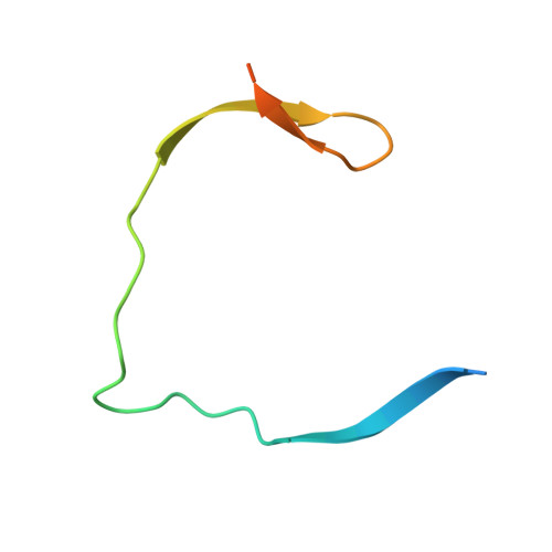
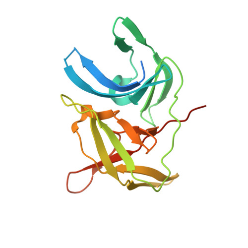
    Three new series of macrocyclic active site-directed inhibitors of the Zika virus (ZIKV) NS2B-NS3 protease were synthesized. First, attempts were made to replace the basic P3 lysine residue of our previously described inhibitors with uncharged and more hydrophobic residues. This provided numerous compounds with inhibition constants between 30 and 50 nM. A stronger reduction of the inhibitory potency was observed when the P2 lysine was replaced by neutral residues, all of these inhibitors possess K i values >1 µM. However, it is possible to replace the P2 lysine with the less basic 3-aminomethylphenylalanine, which provides a similarly potent inhibitor of the ZIKV protease (K i  = 2.69 nM). Crystal structure investigations showed that the P2 benzylamine structure forms comparable interactions with the protease as lysine. Twelve additional structures of these inhibitors in complex with the protease were determined, which explain many, but not all, SAR data obtained in this study. All individual modifications in the P2 or P3 position resulted in inhibitors with low antiviral efficacy in cell culture. Therefore, a third inhibitor series with combined modifications was synthesized; all of them contain a more hydrophobic  d-cyclohexylalanine in the linker segment. At a concentration of 40 µM, two of these compounds possess similar antiviral potency as ribavirin at 100 µM. Due to their reliable crystallization in complex with the ZIKV protease, these cyclic compounds are very well suited for a rational structure-based development of improved inhibitors.


  • Organizational Affiliation
    • Institute of Pharmaceutical Chemistry, Philipps University of Marburg, Marburg, Germany.

Macromolecule Content 

  • Total Structure Weight: 25.55 kDa 
  • Atom Count: 1,712 
  • Modeled Residue Count: 187 
  • Deposited Residue Count: 231 
  • Unique protein chains: 2

Macromolecules

Find similar proteins by:|  3D Structure
Entity ID: 1
MoleculeChains  Sequence LengthOrganismDetailsImage
Serine protease subunit NS2B53Zika virusMutation(s): 0 
UniProt
Find proteins for Q32ZE1 (Zika virus)
Explore Q32ZE1 
Go to UniProtKB:  Q32ZE1
Entity Groups
Sequence Clusters30% Identity50% Identity70% Identity90% Identity95% Identity100% Identity
UniProt GroupQ32ZE1
Sequence Annotations
Expand
Reference Sequence
Find similar proteins by:|  3D Structure
Entity ID: 2
MoleculeChains  Sequence LengthOrganismDetailsImage
Serine protease NS3178Zika virusMutation(s): 0 
EC: 3.4.21.91 (PDB Primary Data), 3.6.1.15 (PDB Primary Data), 3.6.4.13 (PDB Primary Data)
UniProt
Find proteins for Q32ZE1 (Zika virus)
Explore Q32ZE1 
Go to UniProtKB:  Q32ZE1
Entity Groups
Sequence Clusters30% Identity50% Identity70% Identity90% Identity95% Identity100% Identity
UniProt GroupQ32ZE1
Sequence Annotations
Expand
Reference Sequence

Small Molecules

Ligands 1 Unique
IDChains Name / Formula / InChI Key2D Diagram3D Interactions
JG0
(Subject of Investigation/LOI)

Query on JG0


Download:Ideal Coordinates CCD File
C [auth B]1-[(8R,15S,18S)-15-(4-azanylbutyl)-18-(2-carbamimidamidoethyl)-4,7,14,17,20-pentakis(oxidanylidene)-3,6,13,16,19-pentazabicyclo[20.3.1]hexacosa-1(25),22(26),23-trien-8-yl]guanidine
C29 H48 N12 O5
BLTNNRHGYHMJIZ-BDTNDASRSA-N

Experimental Data & Validation

Experimental Data

  • Method: X-RAY DIFFRACTION
  • Resolution: 1.38 Å
  • R-Value Free:  0.156 (Depositor), 0.159 (DCC) 
  • R-Value Work:  0.134 (Depositor), 0.136 (DCC) 
  • R-Value Observed: 0.135 (Depositor) 
Space Group: P 21 21 21
Unit Cell:
Length ( Å )Angle ( ˚ )
a = 49.048α = 90
b = 60.617β = 90
c = 82.824γ = 90
Software Package:
Software NamePurpose
PHENIXrefinement
XDSdata reduction
XDSdata scaling
PHASERphasing
Cootmodel building

Structure Validation

View Full Validation Report


Ligand Structure Quality Assessment 


Entry History 

& Funding Information

Deposition Data


Funding OrganizationLocationGrant Number
Not funded--

Revision History  (Full details and data files)

  • Version 1.0: 2023-05-31
    Type: Initial release
  • Version 1.1: 2024-02-07
    Changes: Data collection, Refinement description
  • Version 1.2: 2024-06-12
    Changes: Database references