7ZVC | pdb_00007zvc

Second crystal form of the mature glutamic-class prolyl-endopeptidase neprosin at 1.85 A resolution.


Experimental Data Snapshot

  • Method: X-RAY DIFFRACTION
  • Resolution: 1.85 Å
  • R-Value Free: 
    0.203 (Depositor), 0.188 (DCC) 
  • R-Value Work: 
    0.173 (Depositor) 
  • R-Value Observed: 
    0.174 (Depositor) 

Starting Model: experimental
View more details

wwPDB Validation 3D Report Full Report

Validation slider image for 7ZVC

Ligand Structure Quality Assessment 


This is version 1.2 of the entry. See complete history

Literature

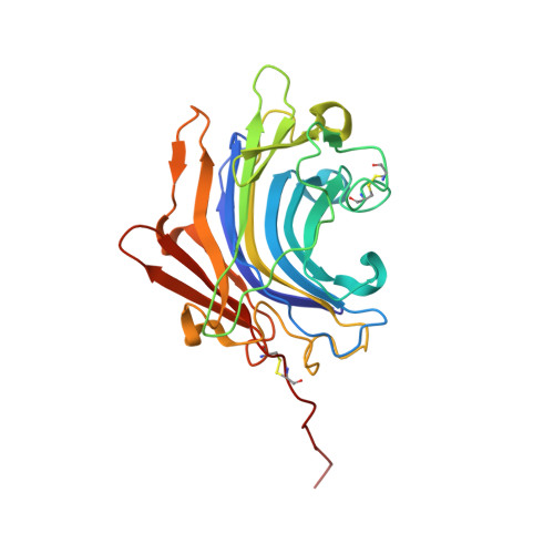
Molecular and in vivo studies of a glutamate-class prolyl-endopeptidase for coeliac disease therapy.

Del Amo-Maestro, L.Mendes, S.R.Rodriguez-Banqueri, A.Garzon-Flores, L.Girbal, M.Rodriguez-Lagunas, M.J.Guevara, T.Franch, A.Perez-Cano, F.J.Eckhard, U.Gomis-Ruth, F.X.

(2022) Nat Commun 13: 4446-4446

  • DOI: https://doi.org/10.1038/s41467-022-32215-1
  • Primary Citation Related Structures: 
    7ZU8, 7ZVA, 7ZVB, 7ZVC

  • PubMed Abstract: 

    The digestion of gluten generates toxic peptides, among which a highly immunogenic proline-rich 33-mer from wheat α-gliadin, that trigger coeliac disease. Neprosin from the pitcher plant is a reported prolyl endopeptidase. Here, we produce recombinant neprosin and its mutants, and find that full-length neprosin is a zymogen, which is self-activated at gastric pH by the release of an all-β pro-domain via a pH-switch mechanism featuring a lysine plug. The catalytic domain is an atypical 7+8-stranded β-sandwich with an extended active-site cleft containing an unprecedented pair of catalytic glutamates. Neprosin efficiently degrades both gliadin and the 33-mer in vitro under gastric conditions and is reversibly inactivated at pH > 5. Moreover, co-administration of gliadin and the neprosin zymogen at the ratio 500:1 reduces the abundance of the 33-mer in the small intestine of mice by up to 90%. Neprosin therefore founds a family of eukaryotic glutamate endopeptidases that fulfils requisites for a therapeutic glutenase.


  • Organizational Affiliation
    • Proteolysis Laboratory; Department of Structural Biology, Molecular Biology Institute of Barcelona (CSIC), Barcelona Science Park; c/Baldiri Reixac, 15-21, 08028, Barcelona, Catalonia, Spain.

Macromolecule Content 

  • Total Structure Weight: 30.12 kDa 
  • Atom Count: 2,351 
  • Modeled Residue Count: 261 
  • Deposited Residue Count: 265 
  • Unique protein chains: 2

Macromolecules

Find similar proteins by:|  3D Structure
Entity ID: 1
MoleculeChains  Sequence LengthOrganismDetailsImage
C-terminal peptidase261Nepenthes ventricosa x Nepenthes alataMutation(s): 0 
UniProt
Find proteins for A0A1L7NZU4 (Nepenthes alata)
Explore A0A1L7NZU4 
Go to UniProtKB:  A0A1L7NZU4
Entity Groups
Sequence Clusters30% Identity50% Identity70% Identity90% Identity95% Identity100% Identity
UniProt GroupA0A1L7NZU4
Glycosylation
Glycosylation Sites: 2
Sequence Annotations
Expand
Reference Sequence
Find similar proteins by:  Sequence   |   3D Structure  
Entity ID: 2
MoleculeChains  Sequence LengthOrganismDetailsImage
GLY-GLY-GLY-GLYB [auth C]4Nepenthes ventricosa x Nepenthes alataMutation(s): 0 

Oligosaccharides

Help  
Entity ID: 3
MoleculeChains Length2D Diagram GlycosylationD Interactions
beta-D-mannopyranose-(1-4)-2-acetamido-2-deoxy-beta-D-glucopyranose-(1-4)-2-acetamido-2-deoxy-beta-D-glucopyranoseC [auth B]3N-Glycosylation
Glycosylation Resources
GlyTouCan: G15407YE
GlyCosmos: G15407YE
GlyGen: G15407YE

Small Molecules

Ligands 4 Unique
IDChains Name / Formula / InChI Key2D Diagram3D Interactions
NAG

Query on NAG


Download:Ideal Coordinates CCD File
D [auth A]2-acetamido-2-deoxy-beta-D-glucopyranose
C8 H15 N O6
OVRNDRQMDRJTHS-FMDGEEDCSA-N
SO4

Query on SO4


Download:Ideal Coordinates CCD File
E [auth A],
F [auth A],
G [auth A]
SULFATE ION
O4 S
QAOWNCQODCNURD-UHFFFAOYSA-L
GLY

Query on GLY


Download:Ideal Coordinates CCD File
I [auth A]GLYCINE
C2 H5 N O2
DHMQDGOQFOQNFH-UHFFFAOYSA-N
NI

Query on NI


Download:Ideal Coordinates CCD File
H [auth A]NICKEL (II) ION
Ni
VEQPNABPJHWNSG-UHFFFAOYSA-N

Experimental Data & Validation

Experimental Data

  • Method: X-RAY DIFFRACTION
  • Resolution: 1.85 Å
  • R-Value Free:  0.203 (Depositor), 0.188 (DCC) 
  • R-Value Work:  0.173 (Depositor) 
  • R-Value Observed: 0.174 (Depositor) 
Space Group: P 1 21 1
Unit Cell:
Length ( Å )Angle ( ˚ )
a = 58.86α = 90
b = 40.63β = 107.98
c = 64.63γ = 90
Software Package:
Software NamePurpose
XDSdata reduction
XSCALEdata scaling
PHASERphasing
BUSTERrefinement
Cootmodel building

Structure Validation

View Full Validation Report


Ligand Structure Quality Assessment 


Entry History 

& Funding Information

Deposition Data


Funding OrganizationLocationGrant Number
Not funded--

Revision History  (Full details and data files)

  • Version 1.0: 2022-08-10
    Type: Initial release
  • Version 1.1: 2024-01-31
    Changes: Data collection, Refinement description
  • Version 1.2: 2024-11-20
    Changes: Structure summary