7ZTY | pdb_00007zty

Structure of Vps39 N-terminal domain from Chaetomium thermophilum


Experimental Data Snapshot

  • Method: X-RAY DIFFRACTION
  • Resolution: 2.89 Å
  • R-Value Free: 
    0.298 (Depositor), 0.305 (DCC) 
  • R-Value Work: 
    0.269 (Depositor), 0.269 (DCC) 
  • R-Value Observed: 
    0.270 (Depositor) 

Starting Model: in silico
View more details

wwPDB Validation 3D Report Full Report

Validation slider image for 7ZTY

This is version 1.3 of the entry. See complete history

Literature

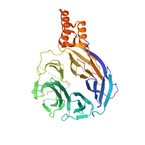
Structure of the HOPS tethering complex, a lysosomal membrane fusion machinery.

Shvarev, D.Schoppe, J.Konig, C.Perz, A.Fullbrunn, N.Kiontke, S.Langemeyer, L.Januliene, D.Schnelle, K.Kummel, D.Frohlich, F.Moeller, A.Ungermann, C.

(2022) Elife 11

  • DOI: https://doi.org/10.7554/eLife.80901
  • Primary Citation Related Structures: 
    7ZTY, 7ZU0

  • PubMed Abstract: 

    Lysosomes are essential for cellular recycling, nutrient signaling, autophagy, and pathogenic bacteria and viruses invasion. Lysosomal fusion is fundamental to cell survival and requires HOPS, a conserved heterohexameric tethering complex. On the membranes to be fused, HOPS binds small membrane-associated GTPases and assembles SNAREs for fusion, but how the complex fulfills its function remained speculative. Here, we used cryo-electron microscopy to reveal the structure of HOPS. Unlike previously reported, significant flexibility of HOPS is confined to its extremities, where GTPase binding occurs. The SNARE-binding module is firmly attached to the core, therefore, ideally positioned between the membranes to catalyze fusion. Our data suggest a model for how HOPS fulfills its dual functionality of tethering and fusion and indicate why it is an essential part of the membrane fusion machinery.


  • Organizational Affiliation
    • Department of Biology/Chemistry, Structural Biology section, Osnabrück University, Osnabrück, Germany.

Macromolecule Content 

  • Total Structure Weight: 54.94 kDa 
  • Atom Count: 2,691 
  • Modeled Residue Count: 345 
  • Deposited Residue Count: 502 
  • Unique protein chains: 1

Macromolecules

Find similar proteins by:|  3D Structure
Entity ID: 1
MoleculeChains  Sequence LengthOrganismDetailsImage
CNH domain-containing protein502Thermochaetoides thermophilaMutation(s): 0 
Gene Names: CTHT_0004950
UniProt
Find proteins for G0RY05 (Chaetomium thermophilum (strain DSM 1495 / CBS 144.50 / IMI 039719))
Explore G0RY05 
Go to UniProtKB:  G0RY05
Entity Groups
Sequence Clusters30% Identity50% Identity70% Identity90% Identity95% Identity100% Identity
UniProt GroupG0RY05
Sequence Annotations
Expand
Reference Sequence

Small Molecules

Modified Residues  1 Unique
IDChains TypeFormula2D DiagramParent
MSE
Query on MSE
A
L-PEPTIDE LINKINGC5 H11 N O2 SeMET

Experimental Data & Validation

Experimental Data

  • Method: X-RAY DIFFRACTION
  • Resolution: 2.89 Å
  • R-Value Free:  0.298 (Depositor), 0.305 (DCC) 
  • R-Value Work:  0.269 (Depositor), 0.269 (DCC) 
  • R-Value Observed: 0.270 (Depositor) 
Space Group: P 21 21 2
Unit Cell:
Length ( Å )Angle ( ˚ )
a = 64.7α = 90
b = 102.75β = 90
c = 59.56γ = 90
Software Package:
Software NamePurpose
PHENIXrefinement
XDSdata reduction
XSCALEdata scaling
PHENIXphasing

Structure Validation

View Full Validation Report


Entry History 

& Funding Information

Deposition Data


Funding OrganizationLocationGrant Number
German Research Foundation (DFG)GermanySFP944-P17

Revision History  (Full details and data files)

  • Version 1.0: 2022-09-21
    Type: Initial release
  • Version 1.1: 2022-09-28
    Changes: Database references
  • Version 1.2: 2024-05-01
    Changes: Data collection, Database references, Refinement description
  • Version 1.3: 2024-11-20
    Changes: Structure summary