7ZTV | pdb_00007ztv

Crystal structure of mutant AR-LBD (F755L) bound to dihydrotestosterone


Experimental Data Snapshot

  • Method: X-RAY DIFFRACTION
  • Resolution: 1.94 Å
  • R-Value Free: 
    0.256 (Depositor), 0.261 (DCC) 
  • R-Value Work: 
    0.216 (Depositor) 
  • R-Value Observed: 
    0.218 (Depositor) 

Starting Model: experimental
View more details

wwPDB Validation 3D Report Full Report

Validation slider image for 7ZTV

Ligand Structure Quality Assessment 


This is version 1.2 of the entry. See complete history

Literature

A hotspot for posttranslational modifications on the androgen receptor dimer interface drives pathology and anti-androgen resistance.

Alegre-Marti, A.Jimenez-Panizo, A.Martinez-Tebar, A.Poulard, C.Peralta-Moreno, M.N.Abella, M.Anton, R.Chinas, M.Eckhard, U.Piulats, J.M.Rojas, A.M.Fernandez-Recio, J.Rubio-Martinez, J.Le Romancer, M.Aytes, A.Fuentes-Prior, P.Estebanez-Perpina, E.

(2023) Sci Adv 9: eade2175-eade2175

  • DOI: https://doi.org/10.1126/sciadv.ade2175
  • Primary Citation Related Structures: 
    7ZTV, 7ZTX, 7ZTZ, 7ZU1, 7ZU2

  • PubMed Abstract: 

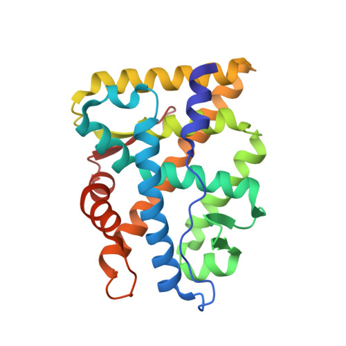
    Mutations of the androgen receptor (AR) associated with prostate cancer and androgen insensitivity syndrome may profoundly influence its structure, protein interaction network, and binding to chromatin, resulting in altered transcription signatures and drug responses. Current structural information fails to explain the effect of pathological mutations on AR structure-function relationship. Here, we have thoroughly studied the effects of selected mutations that span the complete dimer interface of AR ligand-binding domain (AR-LBD) using x-ray crystallography in combination with in vitro, in silico, and cell-based assays. We show that these variants alter AR-dependent transcription and responses to anti-androgens by inducing a previously undescribed allosteric switch in the AR-LBD that increases exposure of a major methylation target, Arg 761 . We also corroborate the relevance of residues Arg 761 and Tyr 764 for AR dimerization and function. Together, our results reveal allosteric coupling of AR dimerization and posttranslational modifications as a disease mechanism with implications for precision medicine.


  • Organizational Affiliation
    • Structural Biology of Nuclear Receptors, Department of Biochemistry and Molecular Biomedicine, Faculty of Biology, University of Barcelona (UB), 08028 Barcelona, Spain.

Macromolecule Content 

  • Total Structure Weight: 29.36 kDa 
  • Atom Count: 2,066 
  • Modeled Residue Count: 243 
  • Deposited Residue Count: 249 
  • Unique protein chains: 1

Macromolecules

Find similar proteins by:|  3D Structure
Entity ID: 1
MoleculeChains  Sequence LengthOrganismDetailsImage
Androgen receptor249Homo sapiensMutation(s): 1 
Gene Names: ArNr3c4
UniProt
Find proteins for P15207 (Rattus norvegicus)
Explore P15207 
Go to UniProtKB:  P15207
Entity Groups
Sequence Clusters30% Identity50% Identity70% Identity90% Identity95% Identity100% Identity
UniProt GroupP15207
Sequence Annotations
Expand
Reference Sequence

Small Molecules

Binding Affinity Annotations 
IDSourceBinding Affinity
DHT BindingDB:  7ZTV Ki: min: 0.2, max: 10 (nM) from 12 assay(s)
Kd: 0.25 (nM) from 1 assay(s)
IC50: min: 0.05, max: 18.5 (nM) from 14 assay(s)
EC50: min: 0.05, max: 20 (nM) from 16 assay(s)

Experimental Data & Validation

Experimental Data

  • Method: X-RAY DIFFRACTION
  • Resolution: 1.94 Å
  • R-Value Free:  0.256 (Depositor), 0.261 (DCC) 
  • R-Value Work:  0.216 (Depositor) 
  • R-Value Observed: 0.218 (Depositor) 
Space Group: P 21 21 21
Unit Cell:
Length ( Å )Angle ( ˚ )
a = 56.175α = 90
b = 65.798β = 90
c = 72.307γ = 90
Software Package:
Software NamePurpose
REFMACrefinement
Aimlessdata scaling
PDB_EXTRACTdata extraction
MOSFLMdata reduction
MOLREPphasing

Structure Validation

View Full Validation Report


Ligand Structure Quality Assessment 


Entry History 

& Funding Information

Deposition Data


Funding OrganizationLocationGrant Number
Not funded--

Revision History  (Full details and data files)

  • Version 1.0: 2023-03-22
    Type: Initial release
  • Version 1.1: 2023-03-29
    Changes: Database references
  • Version 1.2: 2024-02-07
    Changes: Data collection, Refinement description