7ZTL

Crystal structure of a covalently linked Aurora-A N-Myc complex


Experimental Data Snapshot

  • Method: X-RAY DIFFRACTION
  • Resolution: 1.90 Å
  • R-Value Free: 
    0.193 (Depositor), 0.190 (DCC) 
  • R-Value Work: 
    0.163 (Depositor), 0.160 (DCC) 
  • R-Value Observed: 
    0.165 (Depositor) 

Starting Model: experimental
View more details

wwPDB Validation   3D Report Full Report


Ligand Structure Quality Assessment 

Created with Raphaël 2.3.0Worse 01 BetterLigand structure goodness of fit to experimental dataBest fitted JWGClick on this verticalbar to view details

This is version 1.3 of the entry. See complete history


Literature

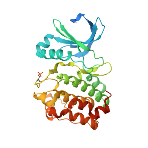
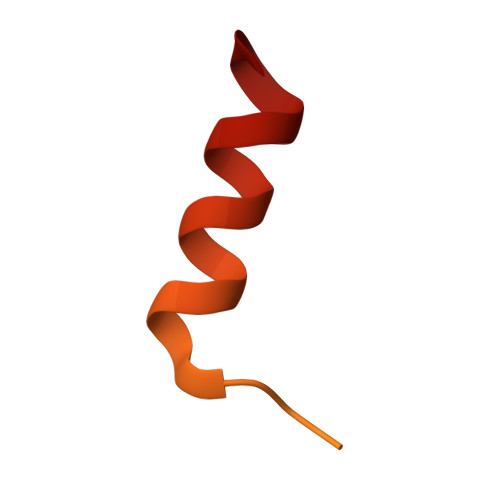
Crystal structure of a covalently linked Aurora-A-MYCN complex.

Diebold, M.Schonemann, L.Eilers, M.Sotriffer, C.Schindelin, H.

(2023) Acta Crystallogr D Struct Biol 79: 1-9

  • DOI: https://doi.org/10.1107/S2059798322011433
  • Primary Citation of Related Structures:  
    7ZTL

  • PubMed Abstract: 

    Formation of the Aurora-A-MYCN complex increases levels of the oncogenic transcription factor MYCN in neuroblastoma cells by abrogating its degradation through the ubiquitin proteasome system. While some small-molecule inhibitors of Aurora-A were shown to destabilize MYCN, clinical trials have not been satisfactory to date. MYCN itself is considered to be `undruggable' due to its large intrinsically disordered regions. Targeting the Aurora-A-MYCN complex rather than Aurora-A or MYCN alone will open new possibilities for drug development and screening campaigns. To overcome the challenges that a ternary system composed of Aurora-A, MYCN and a small molecule entails, a covalently cross-linked construct of the Aurora-A-MYCN complex was designed, expressed and characterized, thus enabling screening and design campaigns to identify selective binders.


  • Organizational Affiliation

    Institute of Pharmacy and Food Chemistry, University of Würzburg, Am Hubland, 97074 Würzburg, Germany.


Macromolecules
Find similar proteins by:  (by identity cutoff)  |  3D Structure
Entity ID: 1
MoleculeChains Sequence LengthOrganismDetailsImage
Aurora kinase A309Homo sapiensMutation(s): 3 
Gene Names: AURKAAIKAIRK1ARK1AURAAYK1BTAKIAK1STK15STK6
EC: 2.7.11.1
UniProt & NIH Common Fund Data Resources
Find proteins for O14965 (Homo sapiens)
Explore O14965 
Go to UniProtKB:  O14965
PHAROS:  O14965
GTEx:  ENSG00000087586 
Entity Groups  
Sequence Clusters30% Identity50% Identity70% Identity90% Identity95% Identity100% Identity
UniProt GroupO14965
Sequence Annotations
Expand
  • Reference Sequence
Find similar proteins by:  (by identity cutoff)  |  3D Structure
Entity ID: 2
MoleculeChains Sequence LengthOrganismDetailsImage
N-myc proto-oncogene protein62Homo sapiensMutation(s): 1 
UniProt & NIH Common Fund Data Resources
Find proteins for P04198 (Homo sapiens)
Explore P04198 
Go to UniProtKB:  P04198
PHAROS:  P04198
GTEx:  ENSG00000134323 
Entity Groups  
Sequence Clusters30% Identity50% Identity70% Identity90% Identity95% Identity100% Identity
UniProt GroupP04198
Sequence Annotations
Expand
  • Reference Sequence
Small Molecules
Ligands 6 Unique
IDChains Name / Formula / InChI Key2D Diagram3D Interactions
ADP
Query on ADP

Download Ideal Coordinates CCD File 
C [auth A]ADENOSINE-5'-DIPHOSPHATE
C10 H15 N5 O10 P2
XTWYTFMLZFPYCI-KQYNXXCUSA-N
JWG (Subject of Investigation/LOI)
Query on JWG

Download Ideal Coordinates CCD File 
K [auth B]4-(3-hydroxy-3-oxopropylamino)-4-oxidanylidene-butanoic acid
C7 H11 N O5
NFQWOKKYYQQDCQ-UHFFFAOYSA-N
BCN
Query on BCN

Download Ideal Coordinates CCD File 
H [auth A]BICINE
C6 H13 N O4
FSVCELGFZIQNCK-UHFFFAOYSA-N
GOL
Query on GOL

Download Ideal Coordinates CCD File 
F [auth A],
G [auth A],
I [auth A]
GLYCEROL
C3 H8 O3
PEDCQBHIVMGVHV-UHFFFAOYSA-N
MG
Query on MG

Download Ideal Coordinates CCD File 
D [auth A],
E [auth A]
MAGNESIUM ION
Mg
JLVVSXFLKOJNIY-UHFFFAOYSA-N
NA
Query on NA

Download Ideal Coordinates CCD File 
J [auth A]SODIUM ION
Na
FKNQFGJONOIPTF-UHFFFAOYSA-N
Modified Residues  1 Unique
IDChains TypeFormula2D DiagramParent
TPO
Query on TPO
A
L-PEPTIDE LINKINGC4 H10 N O6 PTHR
Experimental Data & Validation

Experimental Data

  • Method: X-RAY DIFFRACTION
  • Resolution: 1.90 Å
  • R-Value Free:  0.193 (Depositor), 0.190 (DCC) 
  • R-Value Work:  0.163 (Depositor), 0.160 (DCC) 
  • R-Value Observed: 0.165 (Depositor) 
Space Group: P 32 2 1
Unit Cell:
Length ( Å )Angle ( ˚ )
a = 86.614α = 90
b = 86.614β = 90
c = 91.789γ = 120
Software Package:
Software NamePurpose
PHENIXrefinement
PHASERphasing
Aimlessdata scaling
XDSdata reduction

Structure Validation

View Full Validation Report


Ligand Structure Quality Assessment 

Created with Raphaël 2.3.0Worse 01 BetterLigand structure goodness of fit to experimental dataBest fitted JWGClick on this verticalbar to view details

Entry History & Funding Information

Deposition Data


Funding OrganizationLocationGrant Number
German Research Foundation (DFG)GermanyGRK2243

Revision History  (Full details and data files)

  • Version 1.0: 2023-01-18
    Type: Initial release
  • Version 1.1: 2024-01-31
    Changes: Data collection, Refinement description
  • Version 1.2: 2024-04-03
    Changes: Database references
  • Version 1.3: 2024-11-06
    Changes: Structure summary