7ZTD

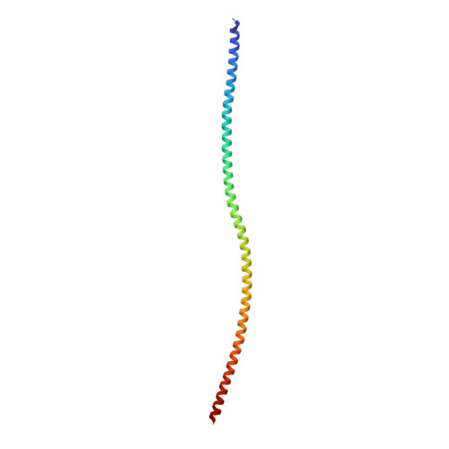
Non-muscle F-actin decorated with non-muscle tropomyosin 3.2


Experimental Data Snapshot

  • Method: ELECTRON MICROSCOPY
  • Resolution: 4.60 Å
  • Aggregation State: FILAMENT 
  • Reconstruction Method: HELICAL 

Starting Models: experimental
View more details

wwPDB Validation   3D Report Full Report


This is version 1.2 of the entry. See complete history


Literature

Structural basis underlying specific biochemical activities of non-muscle tropomyosin isoforms.

Selvaraj, M.Kokate, S.B.Reggiano, G.Kogan, K.Kotila, T.Kremneva, E.DiMaio, F.Lappalainen, P.Huiskonen, J.T.

(2023) Cell Rep 42: 111900-111900

  • DOI: https://doi.org/10.1016/j.celrep.2022.111900
  • Primary Citation of Related Structures:  
    7ZTC, 7ZTD

  • PubMed Abstract: 

    The actin cytoskeleton is critical for cell migration, morphogenesis, endocytosis, organelle dynamics, and cytokinesis. To support diverse cellular processes, actin filaments form a variety of structures with specific architectures and dynamic properties. Key proteins specifying actin filaments are tropomyosins. Non-muscle cells express several functionally non-redundant tropomyosin isoforms, which differentially control the interactions of other proteins, including myosins and ADF/cofilin, with actin filaments. However, the underlying molecular mechanisms have remained elusive. By determining the cryogenic electron microscopy structures of actin filaments decorated by two functionally distinct non-muscle tropomyosin isoforms, Tpm1.6 and Tpm3.2, we reveal that actin filament conformation remains unaffected upon binding. However, Tpm1.6 and Tpm3.2 follow different paths along the actin filament major groove, providing an explanation for their incapability to co-polymerize on actin filaments. We also elucidate the molecular basis underlying specific roles of Tpm1.6 and Tpm3.2 in myosin II activation and protecting actin filaments from ADF/cofilin-catalyzed severing.


  • Organizational Affiliation

    Institute of Biotechnology, Helsinki Institute of Life Science HiLIFE, University of Helsinki, P.O. Box 56, 00014 Helsinki, Finland.


Macromolecules
Find similar proteins by:  (by identity cutoff)  |  3D Structure
Entity ID: 1
MoleculeChains Sequence LengthOrganismDetailsImage
actin, cytoplasmic 1375Homo sapiensMutation(s): 0 
EC: 3.6.4
UniProt & NIH Common Fund Data Resources
Find proteins for P60709 (Homo sapiens)
Explore P60709 
Go to UniProtKB:  P60709
PHAROS:  P60709
GTEx:  ENSG00000075624 
Entity Groups  
Sequence Clusters30% Identity50% Identity70% Identity90% Identity95% Identity100% Identity
UniProt GroupP60709
Sequence Annotations
Expand
  • Reference Sequence
Find similar proteins by:  (by identity cutoff)  |  3D Structure
Entity ID: 2
MoleculeChains Sequence LengthOrganismDetailsImage
Non-muscle tropomyosin 3.2I [auth P],
J [auth Q],
K [auth R],
L [auth S]
135Homo sapiensMutation(s): 0 
Sequence Annotations
Expand
  • Reference Sequence
Small Molecules
Experimental Data & Validation

Experimental Data

  • Method: ELECTRON MICROSCOPY
  • Resolution: 4.60 Å
  • Aggregation State: FILAMENT 
  • Reconstruction Method: HELICAL 
EM Software:
TaskSoftware PackageVersion
MODEL REFINEMENTPHENIX
RECONSTRUCTIONRELION

Structure Validation

View Full Validation Report


Entry History & Funding Information

Deposition Data


Funding OrganizationLocationGrant Number
Academy of FinlandFinland302161
Sigrid Juselius FoundationFinland4708344

Revision History  (Full details and data files)

  • Version 1.0: 2023-01-11
    Type: Initial release
  • Version 1.1: 2023-02-15
    Changes: Database references
  • Version 1.2: 2024-07-24
    Changes: Data collection, Refinement description