


Crystal structure of human Urokinase-type plasminogen activator in complex with bicycle peptide inhibitor UK965
Caregnato, A., Angela, P., Mazzoccato, Y., Frasson, N., Angelini, A., Cendron, L.To be published.
Experimental Data Snapshot
Starting Model: experimental
View more details
Entity ID: 1 | |||||
|---|---|---|---|---|---|
| Molecule | Chains | Sequence Length | Organism | Details | Image |
| Urokinase-type plasminogen activator | 284 | Homo sapiens | Mutation(s): 2 Gene Names: PLAU EC: 3.4.21.73 | 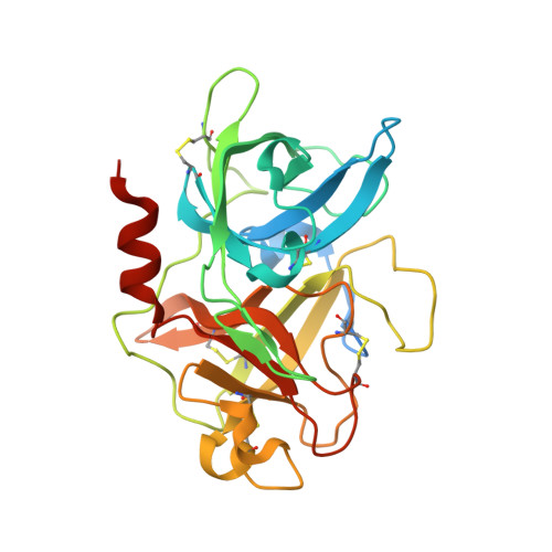 | |
UniProt & NIH Common Fund Data Resources | |||||
PHAROS: P00749 GTEx: ENSG00000122861 | |||||
Entity Groups | |||||
| Sequence Clusters | 30% Identity50% Identity70% Identity90% Identity95% Identity100% Identity | ||||
| UniProt Group | P00749 | ||||
Sequence AnnotationsExpand | |||||
Reference Sequence | |||||
Entity ID: 2 | |||||
|---|---|---|---|---|---|
| Molecule | Chains | Sequence Length | Organism | Details | Image |
| synthetic peptide UK965 | 16 | Homo sapiens | Mutation(s): 0 | 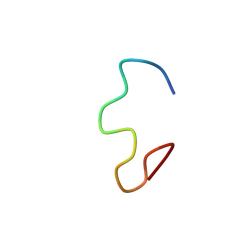 | |
| Ligands 4 Unique | |||||
|---|---|---|---|---|---|
| ID | Chains | Name / Formula / InChI Key | 2D Diagram | 3D Interactions | |
| ZBR (Subject of Investigation/LOI) Download:Ideal Coordinates CCD File | G [auth B] | 1,3,5-tris(bromomethyl)benzene C9 H9 Br3 GHITVUOBZBZMND-UHFFFAOYSA-N |  | ||
| 1PE Download:Ideal Coordinates CCD File | C [auth A] | PENTAETHYLENE GLYCOL C10 H22 O6 JLFNLZLINWHATN-UHFFFAOYSA-N |  | ||
| EDO Download:Ideal Coordinates CCD File | D [auth A], E [auth A] | 1,2-ETHANEDIOL C2 H6 O2 LYCAIKOWRPUZTN-UHFFFAOYSA-N |  | ||
| NH2 (Subject of Investigation/LOI) Download:Ideal Coordinates CCD File | F [auth B] | AMINO GROUP H2 N QGZKDVFQNNGYKY-UHFFFAOYSA-N |  | ||
| Length ( Å ) | Angle ( ˚ ) |
|---|---|
| a = 120.906 | α = 90 |
| b = 120.906 | β = 90 |
| c = 42.716 | γ = 120 |
| Software Name | Purpose |
|---|---|
| REFMAC | refinement |
| PDB_EXTRACT | data extraction |
| XDS | data reduction |
| Aimless | data scaling |
| MOLREP | phasing |
| Funding Organization | Location | Grant Number |
|---|---|---|
| Italian Ministry of Education | Italy | -- |


