7ZPU | pdb_00007zpu

Crystal structure of MreB from Geobacillus stearothermophilus ATCC7953 in complex with ATP


Experimental Data Snapshot

  • Method: X-RAY DIFFRACTION
  • Resolution: 1.96 Å
  • R-Value Free: 
    0.244 (Depositor), 0.246 (DCC) 
  • R-Value Work: 
    0.194 (Depositor), 0.195 (DCC) 
  • R-Value Observed: 
    0.197 (Depositor) 

Starting Model: experimental
View more details

wwPDB Validation 3D Report Full Report

Validation slider image for 7ZPU

Ligand Structure Quality Assessment 


This is version 1.3 of the entry. See complete history

Literature

On the role of nucleotides and lipids in the polymerization of the actin homolog MreB from a Gram-positive bacterium.

Mao, W.Renner, L.D.Cornilleau, C.Li de la Sierra-Gallay, I.Afensiss, S.Benlamara, S.Ah-Seng, Y.Van Tilbeurgh, H.Nessler, S.Bertin, A.Chastanet, A.Carballido-Lopez, R.

(2023) Elife 12

  • DOI: https://doi.org/10.7554/eLife.84505
  • Primary Citation Related Structures: 
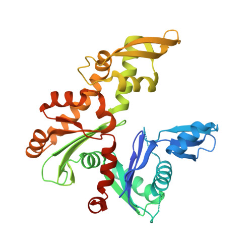
    7ZPT, 7ZPU, 8AZG

  • PubMed Abstract: 

    In vivo, bacterial actin MreB assembles into dynamic membrane-associated filamentous structures that exhibit circumferential motion around the cell. Current knowledge of MreB biochemical and polymerization properties in vitro remains limited and is mostly based on MreB proteins from Gram-negative species. In this study, we report the first observation of organized protofilaments by electron microscopy and the first 3D-structure of MreB from a Gram-positive bacterium. We show that Geobacillus stearothermophilus MreB forms straight pairs of protofilaments on lipid surfaces in the presence of ATP or GTP, but not in the presence of ADP, GDP or non-hydrolysable ATP analogs. We demonstrate that membrane anchoring is mediated by two spatially close short hydrophobic sequences while electrostatic interactions also contribute to lipid binding, and show that the population of membrane-bound protofilament doublets is in steady-state. In solution, protofilament doublets were not detected in any condition tested. Instead, MreB formed large sheets regardless of the bound nucleotide, albeit at a higher critical concentration. Altogether, our results indicate that both lipids and ATP are facilitators of MreB polymerization, and are consistent with a dual effect of ATP hydrolysis, in promoting both membrane binding and filaments assembly/disassembly.


  • Organizational Affiliation
    • Université Paris-Saclay, INRAE, AgroParisTech, Micalis Institute, Jouy-en-Josas, France.

Macromolecule Content 

  • Total Structure Weight: 36.95 kDa 
  • Atom Count: 2,467 
  • Modeled Residue Count: 319 
  • Deposited Residue Count: 340 
  • Unique protein chains: 1

Macromolecules

Find similar proteins by:|  3D Structure
Entity ID: 1
MoleculeChains  Sequence LengthOrganismDetailsImage
Cell shape-determining protein MreBA [auth M]340Geobacillus stearothermophilus ATCC 7953Mutation(s): 0 
Gene Names: mreBB4109_2230B4114_2117GS458_2390
UniProt
Find proteins for A0A150MJ77 (Geobacillus stearothermophilus)
Explore A0A150MJ77 
Go to UniProtKB:  A0A150MJ77
Entity Groups
Sequence Clusters30% Identity50% Identity70% Identity90% Identity95% Identity100% Identity
UniProt GroupA0A150MJ77
Sequence Annotations
Expand
Reference Sequence

Small Molecules

Ligands 1 Unique
IDChains Name / Formula / InChI Key2D Diagram3D Interactions
ATP
(Subject of Investigation/LOI)

Query on ATP


Download:Ideal Coordinates CCD File
B [auth M]ADENOSINE-5'-TRIPHOSPHATE
C10 H16 N5 O13 P3
ZKHQWZAMYRWXGA-KQYNXXCUSA-N

Experimental Data & Validation

Experimental Data

  • Method: X-RAY DIFFRACTION
  • Resolution: 1.96 Å
  • R-Value Free:  0.244 (Depositor), 0.246 (DCC) 
  • R-Value Work:  0.194 (Depositor), 0.195 (DCC) 
  • R-Value Observed: 0.197 (Depositor) 
Space Group: P 1 21 1
Unit Cell:
Length ( Å )Angle ( ˚ )
a = 46.22α = 90
b = 61.6β = 111.53
c = 50.97γ = 90
Software Package:
Software NamePurpose
PHENIXrefinement
XDSdata reduction
XDSdata scaling
MOLREPphasing

Structure Validation

View Full Validation Report


Ligand Structure Quality Assessment 


Entry History 

& Funding Information

Deposition Data


Funding OrganizationLocationGrant Number
French Infrastructure for Integrated Structural Biology (FRISBI)FranceANR-10-INBS-05

Revision History  (Full details and data files)

  • Version 1.0: 2023-05-10
    Type: Initial release
  • Version 1.1: 2023-11-22
    Changes: Data collection, Database references
  • Version 1.2: 2024-02-07
    Changes: Refinement description
  • Version 1.3: 2024-08-14
    Changes: Database references