7ZOY

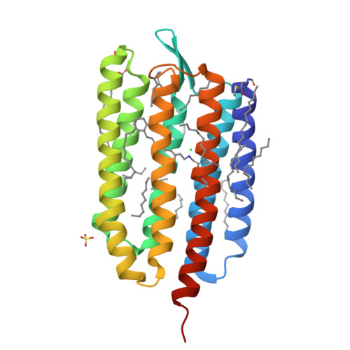
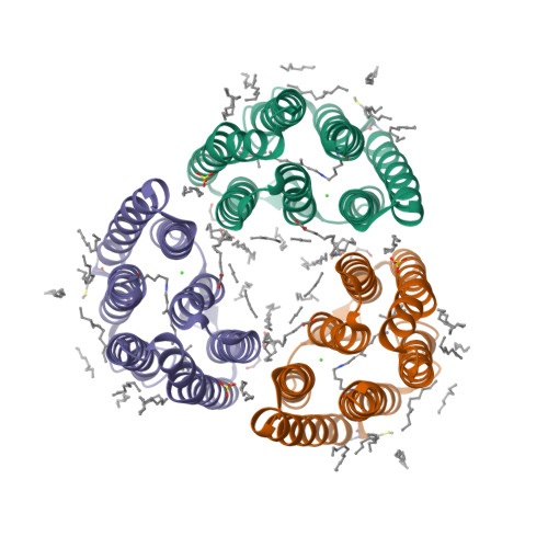
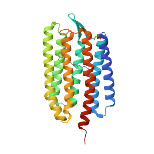
Crystal structure of Synechocystis halorhodopsin (SyHR), SO4-bound form, ground state


Experimental Data Snapshot

  • Method: X-RAY DIFFRACTION
  • Resolution: 1.91 Å
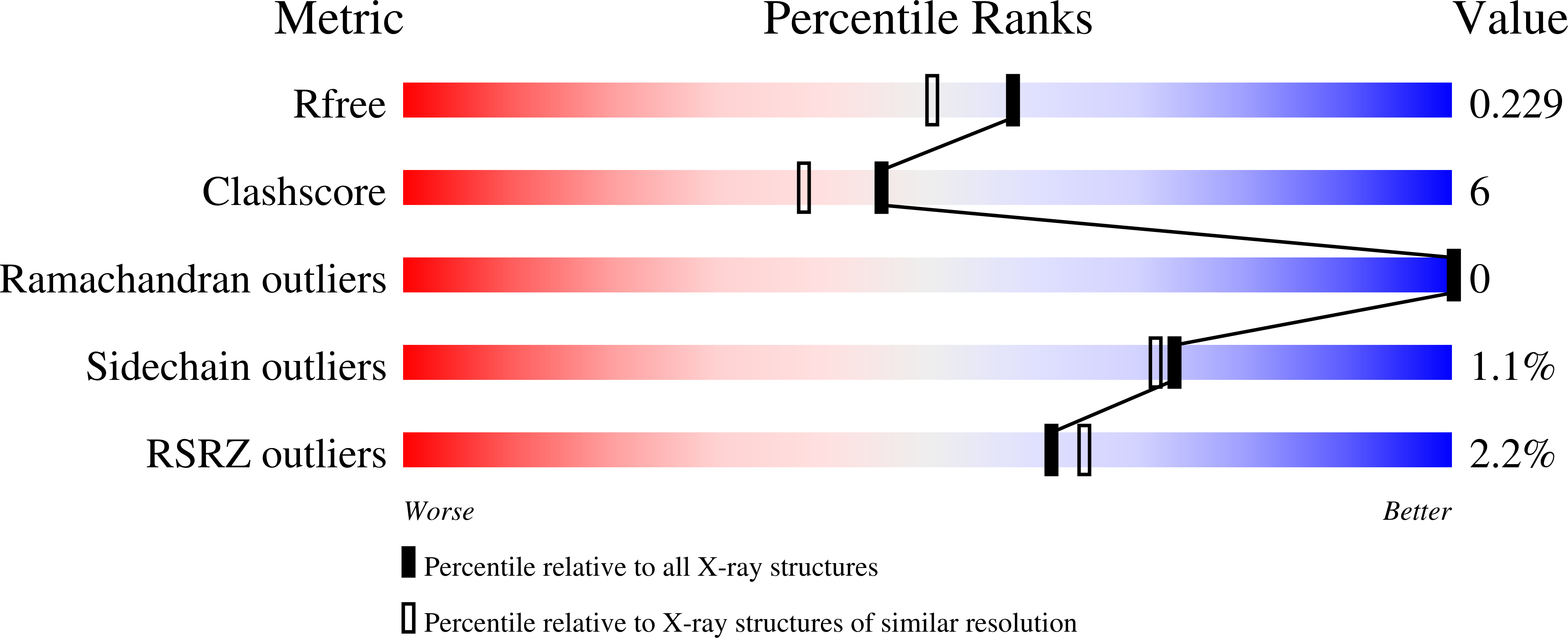
  • R-Value Free: 0.228 
  • R-Value Work: 0.195 
  • R-Value Observed: 0.196 

Starting Model: experimental
View more details

wwPDB Validation   3D Report Full Report


Ligand Structure Quality Assessment 


This is version 1.1 of the entry. See complete history


Literature

Structural insights into light-driven anion pumping in cyanobacteria.

Astashkin, R.Kovalev, K.Bukhdruker, S.Vaganova, S.Kuzmin, A.Alekseev, A.Balandin, T.Zabelskii, D.Gushchin, I.Royant, A.Volkov, D.Bourenkov, G.Koonin, E.Engelhard, M.Bamberg, E.Gordeliy, V.

(2022) Nat Commun 13: 6460-6460

  • DOI: https://doi.org/10.1038/s41467-022-34019-9
  • Primary Citation of Related Structures:  
    7ZOU, 7ZOV, 7ZOW, 7ZOY

  • PubMed Abstract: 

    Transmembrane ion transport is a key process in living cells. Active transport of ions is carried out by various ion transporters including microbial rhodopsins (MRs). MRs perform diverse functions such as active and passive ion transport, photo-sensing, and others. In particular, MRs can pump various monovalent ions like Na + , K + , Cl - , I - , NO 3 - . The only characterized MR proposed to pump sulfate in addition to halides belongs to the cyanobacterium Synechocystis sp. PCC 7509 and is named Synechocystis halorhodopsin (SyHR). The structural study of SyHR may help to understand what makes an MR pump divalent ions. Here we present the crystal structure of SyHR in the ground state, the structure of its sulfate-bound form as well as two photoreaction intermediates, the K and O states. These data reveal the molecular origin of the unique properties of the protein (exceptionally strong chloride binding and proposed pumping of divalent anions) and sheds light on the mechanism of anion release and uptake in cyanobacterial halorhodopsins. The unique properties of SyHR highlight its potential as an optogenetics tool and may help engineer different types of anion pumps with applications in optogenetics.


  • Organizational Affiliation

    Univ. Grenoble Alpes, CEA, CNRS, Institut de Biologie Structurale (IBS), Grenoble, France.


Macromolecules
Find similar proteins by:  (by identity cutoff)  |  3D Structure
Entity ID: 1
MoleculeChains Sequence LengthOrganismDetailsImage
Synechocystis halorhodopsin234Synechocystis sp. PCC 7509Mutation(s): 0 
Entity Groups  
Sequence Clusters30% Identity50% Identity70% Identity90% Identity95% Identity100% Identity
Sequence Annotations
Expand
  • Reference Sequence
Small Molecules
Ligands 5 Unique
IDChains Name / Formula / InChI Key2D Diagram3D Interactions
LFA
Query on LFA

Download Ideal Coordinates CCD File 
D [auth A]
E [auth A]
F [auth A]
G [auth A]
H [auth A]
D [auth A],
E [auth A],
F [auth A],
G [auth A],
H [auth A],
I [auth A],
J [auth A],
K [auth A],
M [auth A],
N [auth A],
O [auth A],
R [auth A],
S [auth A]
EICOSANE
C20 H42
CBFCDTFDPHXCNY-UHFFFAOYSA-N
OLA
Query on OLA

Download Ideal Coordinates CCD File 
L [auth A],
Q [auth A]
OLEIC ACID
C18 H34 O2
ZQPPMHVWECSIRJ-KTKRTIGZSA-N
SO4 (Subject of Investigation/LOI)
Query on SO4

Download Ideal Coordinates CCD File 
C [auth A]SULFATE ION
O4 S
QAOWNCQODCNURD-UHFFFAOYSA-L
GOL
Query on GOL

Download Ideal Coordinates CCD File 
P [auth A]GLYCEROL
C3 H8 O3
PEDCQBHIVMGVHV-UHFFFAOYSA-N
CL
Query on CL

Download Ideal Coordinates CCD File 
B [auth A]CHLORIDE ION
Cl
VEXZGXHMUGYJMC-UHFFFAOYSA-M
Modified Residues  2 Unique
IDChains TypeFormula2D DiagramParent
FME
Query on FME
A
L-PEPTIDE LINKINGC6 H11 N O3 SMET
LYR
Query on LYR
A
L-PEPTIDE LINKINGC26 H42 N2 O2LYS
Experimental Data & Validation

Experimental Data

  • Method: X-RAY DIFFRACTION
  • Resolution: 1.91 Å
  • R-Value Free: 0.228 
  • R-Value Work: 0.195 
  • R-Value Observed: 0.196 
  • Space Group: P 3 2 1
Unit Cell:
Length ( Å )Angle ( ˚ )
a = 62.3α = 90
b = 62.3β = 90
c = 110.14γ = 120
Software Package:
Software NamePurpose
XDSdata reduction
Aimlessdata scaling
MOLREPphasing
PHENIXrefinement

Structure Validation

View Full Validation Report


Ligand Structure Quality Assessment 


Entry History & Funding Information

Deposition Data


Funding OrganizationLocationGrant Number
Agence Nationale de la Recherche (ANR)FranceANR-15-CE11-0029-02
Ministry of Science and Higher Education of the Russian FederationRussian Federation075-00337-20-03/FSMG-2020-0003
French Infrastructure for Integrated Structural Biology (FRISBI)FranceANR-10-INSB-05-02
Grenoble Alliance for Integrated Structural Cell Biology (GRAL)FranceANR-10-LABX-49-01
Russian Science FoundationRussian Federation16-15-00242

Revision History  (Full details and data files)

  • Version 1.0: 2022-11-09
    Type: Initial release
  • Version 1.1: 2024-01-31
    Changes: Data collection, Refinement description