


X-ray structure of the haloalkane dehalogenase HaloTag7 bound to a butyltrifluoromethanesulfonamide tetramethylrhodamine ligand (FSAm(4)-TMR)
Tarnawski, M., Kompa, J., Johnsson, K., Hiblot, J.To be published.
Experimental Data Snapshot
Starting Model: experimental
View more details
Entity ID: 1 | |||||
|---|---|---|---|---|---|
| Molecule | Chains | Sequence Length | Organism | Details | Image |
| Haloalkane dehalogenase | 293 | Rhodococcus sp. (in: high G+C Gram-positive bacteria) | Mutation(s): 21 Gene Names: dhaA EC: 3.8.1.5 | 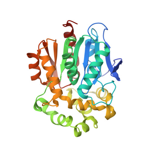 | |
UniProt | |||||
Entity Groups | |||||
| Sequence Clusters | 30% Identity50% Identity70% Identity90% Identity95% Identity100% Identity | ||||
| UniProt Group | P0A3G3 | ||||
Sequence AnnotationsExpand | |||||
Reference Sequence | |||||
| Ligands 1 Unique | |||||
|---|---|---|---|---|---|
| ID | Chains | Name / Formula / InChI Key | 2D Diagram | 3D Interactions | |
| IYE (Subject of Investigation/LOI) Download:Ideal Coordinates CCD File | C [auth A], D [auth B] | [9-[2-carboxy-5-[2-[2-[4-(trifluoromethylsulfonylamino)butoxy]ethoxy]ethylcarbamoyl]phenyl]-6-(dimethylamino)xanthen-3-ylidene]-dimethyl-azanium C34 H40 F3 N4 O8 S CKSQAQHVRBNIHM-UHFFFAOYSA-O |  | ||
| Length ( Å ) | Angle ( ˚ ) |
|---|---|
| a = 44.171 | α = 71.51 |
| b = 49.795 | β = 89.953 |
| c = 78.718 | γ = 67.994 |
| Software Name | Purpose |
|---|---|
| PHENIX | refinement |
| XDS | data reduction |
| XSCALE | data scaling |
| PHASER | phasing |
| STARANISO | data scaling |
| Funding Organization | Location | Grant Number |
|---|---|---|
| Max Planck Society | Germany | -- |


