7ZG0 | pdb_00007zg0

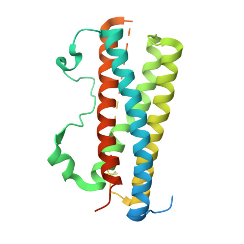
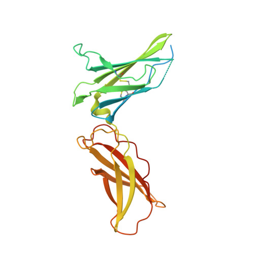
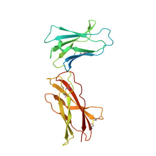
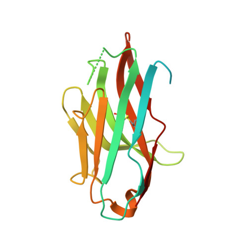
Murine IL-27 in complex with IL-27Ra and a non-competing Nb


Experimental Data Snapshot

  • Method: X-RAY DIFFRACTION
  • Resolution: 3.18 Å
  • R-Value Free: 
    0.278 (Depositor), 0.286 (DCC) 
  • R-Value Work: 
    0.229 (Depositor), 0.257 (DCC) 
  • R-Value Observed: 
    0.232 (Depositor) 

Starting Model: in silico
View more details

wwPDB Validation 3D Report Full Report

Validation slider image for 7ZG0

Ligand Structure Quality Assessment 


This is version 1.2 of the entry. See complete history

Literature

Structural basis of activation and antagonism of receptor signaling mediated by interleukin-27.

Skladanowska, K.Bloch, Y.Strand, J.White, K.F.Hua, J.Aldridge, D.Welin, M.Logan, D.T.Soete, A.Merceron, R.Murphy, C.Provost, M.Bazan, J.F.Hunter, C.A.Hill, J.A.Savvides, S.N.

(2022) Cell Rep 41: 111490-111490

  • DOI: https://doi.org/10.1016/j.celrep.2022.111490
  • Primary Citation Related Structures: 
    7ZG0, 7ZXK

  • PubMed Abstract: 

    Interleukin-27 (IL-27) uniquely assembles p28 and EBI3 subunits to a heterodimeric cytokine that signals via IL-27Rα and gp130. To provide the structural framework for receptor activation by IL-27 and its emerging therapeutic targeting, we report here crystal structures of mouse IL-27 in complex with IL-27Rα and of human IL-27 in complex with SRF388, a monoclonal antibody undergoing clinical trials with oncology indications. One face of the helical p28 subunit interacts with EBI3, while the opposite face nestles into the interdomain elbow of IL-27Rα to juxtapose IL-27Rα to EBI3. This orients IL-27Rα for paired signaling with gp130, which only uses its immunoglobulin domain to bind to IL-27. Such a signaling complex is distinct from those mediated by IL-12 and IL-23. The SRF388 binding epitope on IL-27 overlaps with the IL-27Rα interaction site explaining its potent antagonistic properties. Collectively, our findings will facilitate the mechanistic interrogation, engineering, and therapeutic targeting of IL-27.


  • Organizational Affiliation
    • Unit for Structural Biology, Department of Biochemistry and Microbiology Ghent University, Technologiepark 71, 9052 Ghent, Belgium; Unit for Structural Biology, VIB-UGent Center for Inflammation Research, Technologiepark 71, 9052 Ghent, Belgium.

Macromolecule Content 

  • Total Structure Weight: 195.87 kDa 
  • Atom Count: 10,641 
  • Modeled Residue Count: 1,330 
  • Deposited Residue Count: 1,744 
  • Unique protein chains: 4

Macromolecules

Find similar proteins by:|  3D Structure
Entity ID: 1
MoleculeChains  Sequence LengthOrganismDetailsImage
Interleukin-27 subunit alpha
A, B
246Mus musculusMutation(s): 0 
Gene Names: Il27Il27a
UniProt & NIH Common Fund Data Resources
Find proteins for Q8K3I6 (Mus musculus)
Explore Q8K3I6 
Go to UniProtKB:  Q8K3I6
IMPC:  MGI:2384409
Entity Groups
Sequence Clusters30% Identity50% Identity70% Identity90% Identity95% Identity100% Identity
UniProt GroupQ8K3I6
Glycosylation
Glycosylation Sites: 1Go to GlyGen: Q8K3I6-1
Sequence Annotations
Expand
Reference Sequence
Find similar proteins by:|  3D Structure
Entity ID: 2
MoleculeChains  Sequence LengthOrganismDetailsImage
Interleukin-27 subunit beta
C, D
241Mus musculusMutation(s): 0 
Gene Names: Ebi3Il27b
UniProt
Find proteins for O35228 (Mus musculus)
Explore O35228 
Go to UniProtKB:  O35228
Entity Groups
Sequence Clusters30% Identity50% Identity70% Identity90% Identity95% Identity100% Identity
UniProt GroupO35228
Glycosylation
Glycosylation Sites: 1Go to GlyGen: O35228-1
Sequence Annotations
Expand
Reference Sequence
Find similar proteins by:|  3D Structure
Entity ID: 3
MoleculeChains  Sequence LengthOrganismDetailsImage
Interleukin-27 receptor subunit alpha
E, F
219Mus musculusMutation(s): 0 
Gene Names: Il27raTccrWsx1
UniProt
Find proteins for O70394 (Mus musculus)
Explore O70394 
Go to UniProtKB:  O70394
Entity Groups
Sequence Clusters30% Identity50% Identity70% Identity90% Identity95% Identity100% Identity
UniProt GroupO70394
Glycosylation
Glycosylation Sites: 1Go to GlyGen: O70394-1
Sequence Annotations
Expand
Reference Sequence
Find similar proteins by:|  3D Structure
Entity ID: 4
MoleculeChains  Sequence LengthOrganismDetailsImage
Nanobody 5
G, H
166Lama glamaMutation(s): 0 

Oligosaccharides

Help  
Entity ID: 5
MoleculeChains Length2D Diagram GlycosylationD Interactions
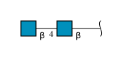
2-acetamido-2-deoxy-beta-D-glucopyranose-(1-4)-2-acetamido-2-deoxy-beta-D-glucopyranose
I, J
2N-Glycosylation
Glycosylation Resources
GlyTouCan: G42666HT
GlyCosmos: G42666HT
GlyGen: G42666HT

Experimental Data & Validation

Experimental Data

  • Method: X-RAY DIFFRACTION
  • Resolution: 3.18 Å
  • R-Value Free:  0.278 (Depositor), 0.286 (DCC) 
  • R-Value Work:  0.229 (Depositor), 0.257 (DCC) 
  • R-Value Observed: 0.232 (Depositor) 
Space Group: C 1 2 1
Unit Cell:
Length ( Å )Angle ( ˚ )
a = 167.242α = 90
b = 137.707β = 117.49
c = 111.479γ = 90
Software Package:
Software NamePurpose
PHENIXrefinement
XDSdata reduction
XDSdata scaling
PHASERphasing

Structure Validation

View Full Validation Report


Ligand Structure Quality Assessment 


Entry History 

& Funding Information

Deposition Data


Funding OrganizationLocationGrant Number
Research Foundation - Flanders (FWO)BelgiumG0E1516N
Research Foundation - Flanders (FWO)BelgiumG0B4918N
Research Foundation - Flanders (FWO)Belgium12S0519N
H2020 Marie Curie Actions of the European CommissionEuropean Union--
Research Foundation - Flanders (FWO)BelgiumHercules

Revision History  (Full details and data files)

  • Version 1.0: 2022-11-02
    Type: Initial release
  • Version 1.1: 2024-05-01
    Changes: Data collection, Refinement description
  • Version 1.2: 2024-10-16
    Changes: Structure summary