7ZBO | pdb_00007zbo

Amine Dehydrogenase MATOUAmDH2 in complex with NADP+


Experimental Data Snapshot

  • Method: X-RAY DIFFRACTION
  • Resolution: 2.32 Å
  • R-Value Free: 
    0.288 (Depositor), 0.289 (DCC) 
  • R-Value Work: 
    0.237 (Depositor), 0.241 (DCC) 
  • R-Value Observed: 
    0.240 (Depositor) 

Starting Model: experimental
View more details

wwPDB Validation 3D Report Full Report

Validation slider image for 7ZBO

Ligand Structure Quality Assessment 


This is version 1.4 of the entry. See complete history

Literature

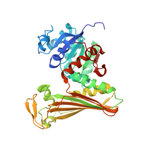
Structure and Mutation of the Native Amine Dehydrogenase MATOUAmDH2.

Bennett, M.Ducrot, L.Vergne-Vaxelaire, C.Grogan, G.

(2022) Chembiochem 23: e202200136-e202200136

  • DOI: https://doi.org/10.1002/cbic.202200136
  • Primary Citation Related Structures: 
    7R09, 7ZBO

  • PubMed Abstract: 

    Native amine dehydrogenases (nat-AmDHs) have recently emerged as a potentially valuable new reservoir of enzymes for the sustainable and selective synthesis of chiral amines, catalyzing the NAD(P)H-dependent ammoniation of carbonyl compounds with high activity and selectivity. MATOUAmDH2, recently identified from the Marine Atlas of Tara Oceans Unigenes (MATOUv1) database of eukaryotic genes, displays exceptional catalytic performance against its best identified substrate, isobutyraldehyde, as well as having broader substrate scope than other nat-AmDHs. In the interests of providing a platform for the rational engineering of this and other nat-AmDHs, we have determined the structure of MATOUAmDH2 in complex with NADP + and also with the cofactor and cyclohexylamine. Monomers within the structure are representative of more open and closed conformations of the enzyme and illustrate the profound changes undergone by nat-AmDHs during the catalytic cycle. An alanine screen of active site residues revealed that M215A and L180A are more active than the wild-type enzyme for the amination of cyclohexanone with ammonia and methylamine respectively; the latter suggests that AmDHs have the potential to be engineered for the improved production of secondary amines.


  • Organizational Affiliation
    • Department of Chemistry, University of York, Heslington, York YO10 5DD, UK.

Macromolecule Content 

  • Total Structure Weight: 155.97 kDa 
  • Atom Count: 10,633 
  • Modeled Residue Count: 1,371 
  • Deposited Residue Count: 1,404 
  • Unique protein chains: 1

Macromolecules

Find similar proteins by:|  3D Structure
Entity ID: 1
MoleculeChains  Sequence LengthOrganismDetailsImage
Amine Dehydrogenase
A, B, C, D
351metagenomeMutation(s): 0 

Experimental Data & Validation

Experimental Data

  • Method: X-RAY DIFFRACTION
  • Resolution: 2.32 Å
  • R-Value Free:  0.288 (Depositor), 0.289 (DCC) 
  • R-Value Work:  0.237 (Depositor), 0.241 (DCC) 
  • R-Value Observed: 0.240 (Depositor) 
Space Group: P 21 21 21
Unit Cell:
Length ( Å )Angle ( ˚ )
a = 91.098α = 90
b = 112.195β = 90
c = 153.146γ = 90
Software Package:
Software NamePurpose
REFMACrefinement
PDB_EXTRACTdata extraction
XDSdata reduction
SCALAdata scaling
MOLREPphasing

Structure Validation

View Full Validation Report


Ligand Structure Quality Assessment 


Entry History 

& Funding Information

Deposition Data


Funding OrganizationLocationGrant Number
Not funded--

Revision History  (Full details and data files)

  • Version 1.0: 2022-04-06
    Type: Initial release
  • Version 1.1: 2022-04-13
    Changes: Database references
  • Version 1.2: 2022-04-20
    Changes: Database references
  • Version 1.3: 2022-06-01
    Changes: Database references
  • Version 1.4: 2024-01-31
    Changes: Data collection, Refinement description