7ZBF | pdb_00007zbf

Crystal structure of native Iripin-4 serpin from tick Ixodes ricinus


Experimental Data Snapshot

  • Method: X-RAY DIFFRACTION
  • Resolution: 2.30 Å
  • R-Value Free: 
    0.251 (Depositor), 0.254 (DCC) 
  • R-Value Work: 
    0.197 (Depositor), 0.203 (DCC) 
  • R-Value Observed: 
    0.200 (Depositor) 

Starting Model: experimental
View more details

wwPDB Validation 3D Report Full Report

Validation slider image for 7ZBF

This is version 1.3 of the entry. See complete history

Literature

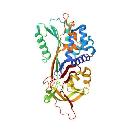
Conformational transition of the Ixodes ricinus salivary serpin Iripin-4.

Kascakova, B.Kotal, J.Havlickova, P.Vopatkova, V.Prudnikova, T.Grinkevich, P.Kuty, M.Chmelar, J.Kuta Smatanova, I.

(2023) Acta Crystallogr D Struct Biol 79: 409-419

  • DOI: https://doi.org/10.1107/S2059798323002322
  • Primary Citation Related Structures: 
    7ZAS, 7ZBF

  • PubMed Abstract: 

    Iripin-4, one of the many salivary serpins from Ixodes ricinus ticks with an as-yet unexplained function, crystallized in two different structural conformations, namely the native partially relaxed state and the cleaved serpin. The native structure was solved at a resolution of 2.3 Å and the structure of the cleaved conformation was solved at 2.0 Å resolution. Furthermore, structural changes were observed when the reactive-centre loop transitioned from the native conformation to the cleaved conformation. In addition to this finding, it was confirmed that Glu341 represents a primary substrate-recognition site for the inhibitory mechanism. The presence of glutamate instead of the typical arginine in the P1 recognition site of all structurally characterized I. ricinus serpins (PDB entries 7b2t, 7pmu and 7ahp), except for the tyrosine in the P1 site of Iripin-2 (formerly IRS-2; PDB entry 3nda), would explain the absence of inhibition of the tested proteases that cleave their substrate after arginine. Further research on Iripin-4 should focus on functional analysis of this interesting serpin.


  • Organizational Affiliation
    • Department of Chemistry, University of South Bohemia in České Budějovice, 370 05 České Budějovice, Czech Republic.

Macromolecule Content 

  • Total Structure Weight: 42.05 kDa 
  • Atom Count: 3,168 
  • Modeled Residue Count: 376 
  • Deposited Residue Count: 376 
  • Unique protein chains: 1

Macromolecules

Find similar proteins by:|  3D Structure
Entity ID: 1
MoleculeChains  Sequence LengthOrganismDetailsImage
Iripin-4 serpin376Ixodes ricinusMutation(s): 3 
UniProt
Find proteins for A0A0K8RJV9 (Ixodes ricinus)
Explore A0A0K8RJV9 
Go to UniProtKB:  A0A0K8RJV9
Entity Groups
Sequence Clusters30% Identity50% Identity70% Identity90% Identity95% Identity100% Identity
UniProt GroupA0A0K8RJV9
Sequence Annotations
Expand
Reference Sequence

Small Molecules

Ligands 1 Unique
IDChains Name / Formula / InChI Key2D Diagram3D Interactions
NI

Query on NI


Download:Ideal Coordinates CCD File
B [auth A]NICKEL (II) ION
Ni
VEQPNABPJHWNSG-UHFFFAOYSA-N

Experimental Data & Validation

Experimental Data

  • Method: X-RAY DIFFRACTION
  • Resolution: 2.30 Å
  • R-Value Free:  0.251 (Depositor), 0.254 (DCC) 
  • R-Value Work:  0.197 (Depositor), 0.203 (DCC) 
  • R-Value Observed: 0.200 (Depositor) 
Space Group: P 31 2 1
Unit Cell:
Length ( Å )Angle ( ˚ )
a = 78.932α = 90
b = 78.932β = 90
c = 117.78γ = 120
Software Package:
Software NamePurpose
XDSdata reduction
XSCALEdata scaling
MOLREPphasing
REFMACrefinement
PDB_EXTRACTdata extraction

Structure Validation

View Full Validation Report


Entry History 

& Funding Information

Deposition Data


Funding OrganizationLocationGrant Number
European Regional Development FundEuropean UnionCZ.02.1.01/0.0/0.0/15_003/0000441
Grant Agency of the Czech RepublicCzech Republic19-14704Y

Revision History  (Full details and data files)

  • Version 1.0: 2023-04-05
    Type: Initial release
  • Version 1.1: 2023-05-03
    Changes: Database references
  • Version 1.2: 2023-05-17
    Changes: Database references
  • Version 1.3: 2024-02-07
    Changes: Data collection, Refinement description