7ZAN | pdb_00007zan

Crystal Structure of human IL-17A in complex with IL-17RA and IL-17RC


Experimental Data Snapshot

  • Method: X-RAY DIFFRACTION
  • Resolution: 5.06 Å
  • R-Value Free: 
    0.353 (Depositor), 0.387 (DCC) 
  • R-Value Work: 
    0.294 (Depositor), 0.317 (DCC) 
  • R-Value Observed: 
    0.297 (Depositor) 

Starting Models: experimental
View more details

wwPDB Validation 3D Report Full Report

Validation slider image for 7ZAN

Ligand Structure Quality Assessment 


This is version 1.3 of the entry. See complete history

Literature

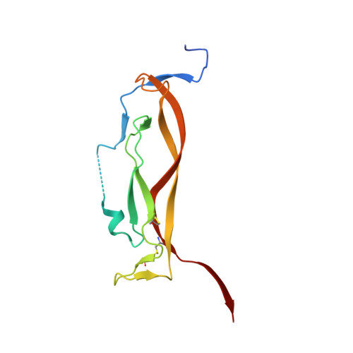
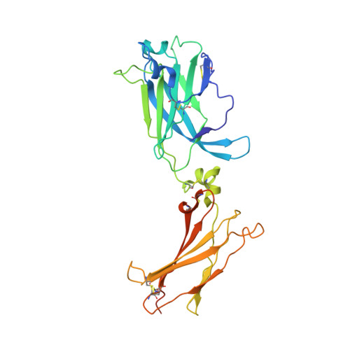
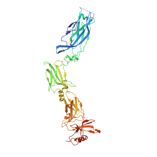
IL-17-induced dimerization of IL-17RA drives the formation of the IL-17 signalosome to potentiate signaling.

Goepfert, A.Barske, C.Lehmann, S.Wirth, E.Willemsen, J.Gudjonsson, J.E.Ward, N.L.Sarkar, M.K.Hemmig, R.Kolbinger, F.Rondeau, J.M.

(2022) Cell Rep 41: 111489-111489

  • DOI: https://doi.org/10.1016/j.celrep.2022.111489
  • Primary Citation Related Structures: 
    5N9B, 7ZAN

  • PubMed Abstract: 

    Signaling through innate immune receptors such as the Toll-like receptor (TLR)/interleukin-1 receptor (IL-1R) superfamily proceeds via the assembly of large membrane-proximal complexes or "signalosomes." Although structurally distinct, the IL-17 receptor family triggers cellular responses that are typical of innate immune receptors. The IL-17RA receptor subunit is shared by several members of the IL-17 family. Using a combination of crystallographic, biophysical, and mutational studies, we show that IL-17A, IL-17F, and IL-17A/F induce IL-17RA dimerization. X-ray analysis of the heteromeric IL-17A complex with the extracellular domains of the IL-17RA and IL-17RC receptors reveals that cytokine-induced IL-17RA dimerization leads to the formation of a 2:2:2 hexameric signaling assembly. Furthermore, we demonstrate that the formation of the IL-17 signalosome potentiates IL-17-induced IL-36γ and CXCL1 mRNA expression in human keratinocytes, compared with a dimerization-defective IL-17RA variant.


  • Organizational Affiliation
    • Novartis Institutes for BioMedical Research, Novartis Pharma AG, 4002 Basel, Switzerland.

Macromolecule Content 

  • Total Structure Weight: 113.87 kDa 
  • Atom Count: 7,417 
  • Modeled Residue Count: 921 
  • Deposited Residue Count: 991 
  • Unique protein chains: 3

Macromolecules

Find similar proteins by:|  3D Structure
Entity ID: 1
MoleculeChains  Sequence LengthOrganismDetailsImage
Interleukin-17A
A, B
123Homo sapiensMutation(s): 2 
Gene Names: IL17ACTLA8IL17
UniProt & NIH Common Fund Data Resources
Find proteins for Q16552 (Homo sapiens)
Explore Q16552 
Go to UniProtKB:  Q16552
PHAROS:  Q16552
GTEx:  ENSG00000112115 
Entity Groups
Sequence Clusters30% Identity50% Identity70% Identity90% Identity95% Identity100% Identity
UniProt GroupQ16552
Sequence Annotations
Expand
Reference Sequence
Find similar proteins by:|  3D Structure
Entity ID: 2
MoleculeChains  Sequence LengthOrganismDetailsImage
Interleukin-17 receptor A292Homo sapiensMutation(s): 3 
Gene Names: IL17RAIL17R
UniProt & NIH Common Fund Data Resources
Find proteins for Q96F46 (Homo sapiens)
Explore Q96F46 
Go to UniProtKB:  Q96F46
PHAROS:  Q96F46
GTEx:  ENSG00000177663 
Entity Groups
Sequence Clusters30% Identity50% Identity70% Identity90% Identity95% Identity100% Identity
UniProt GroupQ96F46
Glycosylation
Glycosylation Sites: 1Go to GlyGen: Q96F46-1
Sequence Annotations
Expand
Reference Sequence
Find similar proteins by:|  3D Structure
Entity ID: 3
MoleculeChains  Sequence LengthOrganismDetailsImage
Isoform 2 of Interleukin-17 receptor C453Homo sapiensMutation(s): 0 
Gene Names: IL17RCUNQ6118/PRO20040/PRO38901
UniProt & NIH Common Fund Data Resources
Find proteins for Q8NAC3 (Homo sapiens)
Explore Q8NAC3 
Go to UniProtKB:  Q8NAC3
PHAROS:  Q8NAC3
GTEx:  ENSG00000163702 
Entity Groups
Sequence Clusters30% Identity50% Identity70% Identity90% Identity95% Identity100% Identity
UniProt GroupQ8NAC3
Glycosylation
Glycosylation Sites: 3Go to GlyGen: Q8NAC3-1
Sequence Annotations
Expand
Reference Sequence

Experimental Data & Validation

Experimental Data

  • Method: X-RAY DIFFRACTION
  • Resolution: 5.06 Å
  • R-Value Free:  0.353 (Depositor), 0.387 (DCC) 
  • R-Value Work:  0.294 (Depositor), 0.317 (DCC) 
  • R-Value Observed: 0.297 (Depositor) 
Space Group: P 65 2 2
Unit Cell:
Length ( Å )Angle ( ˚ )
a = 186.305α = 90
b = 186.305β = 90
c = 238.305γ = 120
Software Package:
Software NamePurpose
BUSTERrefinement
XDSdata reduction
Aimlessdata scaling
PHASERphasing
PDB_EXTRACTdata extraction

Structure Validation

View Full Validation Report


Ligand Structure Quality Assessment 


Entry History 

& Funding Information

Deposition Data


Funding OrganizationLocationGrant Number
Not funded--

Revision History  (Full details and data files)

  • Version 1.0: 2022-10-12
    Type: Initial release
  • Version 1.1: 2022-11-02
    Changes: Database references
  • Version 1.2: 2024-01-31
    Changes: Data collection, Refinement description
  • Version 1.3: 2024-10-16
    Changes: Structure summary