7Z70 | pdb_00007z70

Crystal structure of Angiotensin-1 converting enzyme C-domain in complex with fosinoprilat


Experimental Data Snapshot

  • Method: X-RAY DIFFRACTION
  • Resolution: 1.85 Å
  • R-Value Free: 
    0.213 (Depositor), 0.201 (DCC) 
  • R-Value Work: 
    0.174 (Depositor) 
  • R-Value Observed: 
    0.175 (Depositor) 

Starting Model: experimental
View more details

wwPDB Validation 3D Report Full Report

Validation slider image for 7Z70

Ligand Structure Quality Assessment 


This is version 1.3 of the entry. See complete history

Literature

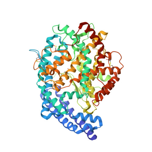
Structural basis for the inhibition of human angiotensin-1 converting enzyme by fosinoprilat.

Cozier, G.E.Newby, E.C.Schwager, S.L.U.Isaac, R.E.Sturrock, E.D.Acharya, K.R.

(2022) FEBS J 289: 6659-6671

  • DOI: https://doi.org/10.1111/febs.16543
  • Primary Citation Related Structures: 
    7Z6Z, 7Z70

  • PubMed Abstract: 

    Human angiotensin I-converting enzyme (ACE) has two isoforms, somatic ACE (sACE) and testis ACE (tACE). The functions of sACE are widespread, with its involvement in blood pressure regulation most extensively studied. sACE is composed of an N-domain (nACE) and a C-domain (cACE), both catalytically active but have significant structural differences, resulting in different substrate specificities. Even though ACE inhibitors are used clinically, they need much improvement because of serious side effects seen in patients (~ 25-30%) with long-term treatment due to nonselective inhibition of nACE and cACE. Investigation into the distinguishing structural features of each domain is therefore of vital importance for the development of domain-specific inhibitors with minimal side effects. Here, we report kinetic data and high-resolution crystal structures of both nACE (1.75 Å) and cACE (1.85 Å) in complex with fosinoprilat, a clinically used inhibitor. These structures allowed detailed analysis of the molecular features conferring domain selectivity by fosinoprilat. Particularly, altered hydrophobic interactions were observed to be a contributing factor. These experimental data contribute to improved understanding of the structural features that dictate ACE inhibitor domain selectivity, allowing further progress towards designing novel 2nd-generation domain-specific potent ACE inhibitors suitable for clinical administration, with a variety of potential future therapeutic benefits. DATABASE: The atomic coordinates and structure factors for nACE-fosinoprilat and cACE-fosinoprilat structures have been deposited with codes 7Z6Z and 7Z70, respectively, in the RCSB Protein Data Bank, www.pdb.org.


  • Organizational Affiliation
    • Department of Biology and Biochemistry, University of Bath, UK.

Macromolecule Content 

  • Total Structure Weight: 71 kDa 
  • Atom Count: 5,469 
  • Modeled Residue Count: 575 
  • Deposited Residue Count: 597 
  • Unique protein chains: 1

Macromolecules

Find similar proteins by:|  3D Structure
Entity ID: 1
MoleculeChains  Sequence LengthOrganismDetailsImage
Angiotensin-converting enzyme597Homo sapiensMutation(s): 5 
Gene Names: ACEDCPDCP1
EC: 3.2.1 (PDB Primary Data), 3.4.15.1 (PDB Primary Data)
UniProt & NIH Common Fund Data Resources
Find proteins for P12821 (Homo sapiens)
Explore P12821 
Go to UniProtKB:  P12821
PHAROS:  P12821
GTEx:  ENSG00000159640 
Entity Groups
Sequence Clusters30% Identity50% Identity70% Identity90% Identity95% Identity100% Identity
UniProt GroupP12821
Glycosylation
Glycosylation Sites: 2Go to GlyGen: P12821-1
Sequence Annotations
Expand
Reference Sequence

Oligosaccharides

Help  
Entity ID: 2
MoleculeChains Length2D Diagram GlycosylationD Interactions
alpha-D-mannopyranose-(1-3)-beta-D-mannopyranose-(1-4)-2-acetamido-2-deoxy-beta-D-glucopyranose-(1-4)-[alpha-L-fucopyranose-(1-6)]2-acetamido-2-deoxy-beta-D-glucopyranose
B
5N-Glycosylation
Glycosylation Resources
GlyTouCan: G42466VF
GlyCosmos: G42466VF
GlyGen: G42466VF

Small Molecules

Ligands 8 Unique
IDChains Name / Formula / InChI Key2D Diagram3D Interactions
KS8
(Subject of Investigation/LOI)

Query on KS8


Download:Ideal Coordinates CCD File
C [auth A]fosinoprilat
C23 H34 N O5 P
WOIWWYDXDVSWAZ-RTWAWAEBSA-N
NAG

Query on NAG


Download:Ideal Coordinates CCD File
D [auth A]2-acetamido-2-deoxy-beta-D-glucopyranose
C8 H15 N O6
OVRNDRQMDRJTHS-FMDGEEDCSA-N
IMD

Query on IMD


Download:Ideal Coordinates CCD File
H [auth A]IMIDAZOLE
C3 H5 N2
RAXXELZNTBOGNW-UHFFFAOYSA-O
ZN

Query on ZN


Download:Ideal Coordinates CCD File
L [auth A]ZINC ION
Zn
PTFCDOFLOPIGGS-UHFFFAOYSA-N
EDO

Query on EDO


Download:Ideal Coordinates CCD File
I [auth A],
J [auth A],
K [auth A]
1,2-ETHANEDIOL
C2 H6 O2
LYCAIKOWRPUZTN-UHFFFAOYSA-N
BO3

Query on BO3


Download:Ideal Coordinates CCD File
E [auth A],
G [auth A]
BORIC ACID
B H3 O3
KGBXLFKZBHKPEV-UHFFFAOYSA-N
ACT

Query on ACT


Download:Ideal Coordinates CCD File
F [auth A]ACETATE ION
C2 H3 O2
QTBSBXVTEAMEQO-UHFFFAOYSA-M
CL

Query on CL


Download:Ideal Coordinates CCD File
M [auth A],
N [auth A]
CHLORIDE ION
Cl
VEXZGXHMUGYJMC-UHFFFAOYSA-M
Modified Residues  1 Unique
IDChains TypeFormula2D DiagramParent
CSO
Query on CSO
A
L-PEPTIDE LINKINGC3 H7 N O3 SCYS
Binding Affinity Annotations 
IDSourceBinding Affinity
KS8 BindingDB:  7Z70 Ki: min: 1.5, max: 1.5 (nM) from 2 assay(s)

Experimental Data & Validation

Experimental Data

  • Method: X-RAY DIFFRACTION
  • Resolution: 1.85 Å
  • R-Value Free:  0.213 (Depositor), 0.201 (DCC) 
  • R-Value Work:  0.174 (Depositor) 
  • R-Value Observed: 0.175 (Depositor) 
Space Group: P 21 21 21
Unit Cell:
Length ( Å )Angle ( ˚ )
a = 56.563α = 90
b = 85.987β = 90
c = 134.686γ = 90
Software Package:
Software NamePurpose
PHENIXrefinement
DIALSdata reduction
Aimlessdata scaling
PHASERphasing

Structure Validation

View Full Validation Report


Ligand Structure Quality Assessment 


Entry History 

& Funding Information

Deposition Data


Funding OrganizationLocationGrant Number
Medical Research Council (MRC, United Kingdom)United KingdomMR/M026647/1

Revision History  (Full details and data files)

  • Version 1.0: 2022-06-22
    Type: Initial release
  • Version 1.1: 2022-11-16
    Changes: Database references
  • Version 1.2: 2024-01-31
    Changes: Data collection, Refinement description
  • Version 1.3: 2024-11-13
    Changes: Structure summary