7Z48 | pdb_00007z48

Bottom part (C5) of bacteriophage SU10 capsid


Experimental Data Snapshot

  • Method: ELECTRON MICROSCOPY
  • Resolution: 4.00 Å
  • Aggregation State: PARTICLE 
  • Reconstruction Method: SINGLE PARTICLE 

wwPDB Validation 3D Report Full Report

Validation slider image for 7Z48

This is version 1.2 of the entry. See complete history

Literature

Tail proteins of phage SU10 reorganize into the nozzle for genome delivery.

Siborova, M.Fuzik, T.Prochazkova, M.Novacek, J.Benesik, M.Nilsson, A.S.Plevka, P.

(2022) Nat Commun 13: 5622-5622

  • DOI: https://doi.org/10.1038/s41467-022-33305-w
  • Primary Citation Related Structures: 
    7Z44, 7Z45, 7Z46, 7Z47, 7Z48, 7Z49, 7Z4A, 7Z4B, 7Z4F

  • PubMed Abstract: 

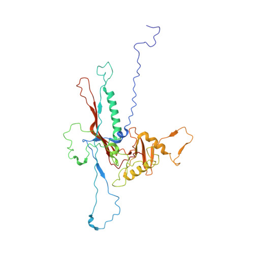
    Escherichia coli phage SU10 belongs to the genus Kuravirus from the class Caudoviricetes of phages with short non-contractile tails. In contrast to other short-tailed phages, the tails of Kuraviruses elongate upon cell attachment. Here we show that the virion of SU10 has a prolate head, containing genome and ejection proteins, and a tail, which is formed of portal, adaptor, nozzle, and tail needle proteins and decorated with long and short fibers. The binding of the long tail fibers to the receptors in the outer bacterial membrane induces the straightening of nozzle proteins and rotation of short tail fibers. After the re-arrangement, the nozzle proteins and short tail fibers alternate to form a nozzle that extends the tail by 28 nm. Subsequently, the tail needle detaches from the nozzle proteins and five types of ejection proteins are released from the SU10 head. The nozzle with the putative extension formed by the ejection proteins enables the delivery of the SU10 genome into the bacterial cytoplasm. It is likely that this mechanism of genome delivery, involving the formation of the tail nozzle, is employed by all Kuraviruses.


  • Organizational Affiliation
    • Central European Institute of Technology, Kamenice 753/5, 625 00, Brno, Czech Republic.

Macromolecule Content 

  • Total Structure Weight: 656.48 kDa 
  • Atom Count: 45,123 
  • Modeled Residue Count: 5,831 
  • Deposited Residue Count: 5,984 
  • Unique protein chains: 1

Macromolecules

Find similar proteins by:|  3D Structure
Entity ID: 1
MoleculeChains  Sequence LengthOrganismDetailsImage
Major head protein352Escherichia phage vB_EcoP_SU10Mutation(s): 0 
UniProt
Find proteins for A0A0B4N1Q7 (Escherichia phage vB_EcoP_SU10)
Explore A0A0B4N1Q7 
Go to UniProtKB:  A0A0B4N1Q7
Entity Groups
Sequence Clusters30% Identity50% Identity70% Identity90% Identity95% Identity100% Identity
UniProt GroupA0A0B4N1Q7
Sequence Annotations
Expand
Reference Sequence

Experimental Data & Validation

Experimental Data

  • Method: ELECTRON MICROSCOPY
  • Resolution: 4.00 Å
  • Aggregation State: PARTICLE 
  • Reconstruction Method: SINGLE PARTICLE 
EM Software:
TaskSoftware PackageVersion
MODEL REFINEMENTPHENIX
MODEL REFINEMENTISOLDE
RECONSTRUCTIONRELION3.1

Structure Validation

View Full Validation Report


Entry History 

& Funding Information

Deposition Data


Funding OrganizationLocationGrant Number
Ministry of Education, Youth and Sports of the Czech RepublicCzech RepublicLL1906

Revision History  (Full details and data files)

  • Version 1.0: 2022-08-10
    Type: Initial release
  • Version 1.1: 2022-10-12
    Changes: Database references
  • Version 1.2: 2024-07-17
    Changes: Data collection