7Z3J | pdb_00007z3j

Structure of crystallisable rat Phospholipase C gamma 1 in complex with inositol 1,4,5-trisphosphate


Experimental Data Snapshot

  • Method: X-RAY DIFFRACTION
  • Resolution: 2.00 Å
  • R-Value Free: 
    0.256 (Depositor), 0.257 (DCC) 
  • R-Value Work: 
    0.224 (Depositor), 0.224 (DCC) 
  • R-Value Observed: 
    0.225 (Depositor) 

Starting Model: experimental
View more details

wwPDB Validation 3D Report Full Report

Validation slider image for 7Z3J

Ligand Structure Quality Assessment 


This is version 1.2 of the entry. See complete history

Literature

Characterization of the membrane interactions of phospholipase C gamma reveals key features of the active enzyme.

Le Huray, K.I.P.Bunney, T.D.Pinotsis, N.Kalli, A.C.Katan, M.

(2022) Sci Adv 8: eabp9688-eabp9688

  • DOI: https://doi.org/10.1126/sciadv.abp9688
  • Primary Citation Related Structures: 
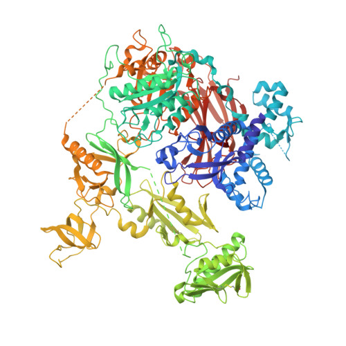
    7Z3J

  • PubMed Abstract: 

    PLCγ enzymes are autoinhibited in resting cells and form key components of intracellular signaling that are also linked to disease development. Insights into physiological and aberrant activation of PLCγ require understanding of an active, membrane-bound form, which can hydrolyze inositol-lipid substrates. Here, we demonstrate that PLCγ1 cannot bind membranes unless the autoinhibition is disrupted. Through extensive molecular dynamics simulations and experimental evidence, we characterize membrane binding by the catalytic core domains and reveal previously unknown sites of lipid interaction. The identified sites act in synergy, overlap with autoinhibitory interfaces, and are shown to be critical for the phospholipase activity in cells. This work provides direct evidence that PLCγ1 is inhibited through obstruction of its membrane-binding surfaces by the regulatory region and that activation must shift PLCγ1 to a conformation competent for membrane binding. Knowledge of the critical sites of membrane interaction extends the mechanistic framework for activation, dysregulation, and therapeutic intervention.


  • Organizational Affiliation
    • Astbury Centre for Structural Molecular Biology, Faculty of Biological Sciences, University of Leeds, Leeds LS2 9JT, UK.

Macromolecule Content 

  • Total Structure Weight: 136.77 kDa 
  • Atom Count: 9,395 
  • Modeled Residue Count: 1,119 
  • Deposited Residue Count: 1,176 
  • Unique protein chains: 1

Macromolecules

Find similar proteins by:|  3D Structure
Entity ID: 1
MoleculeChains  Sequence LengthOrganismDetailsImage
1-phosphatidylinositol 4,5-bisphosphate phosphodiesterase gamma-11,176Rattus norvegicusMutation(s): 0 
Gene Names: Plcg1
EC: 3.1.4.11
UniProt
Find proteins for P10686 (Rattus norvegicus)
Explore P10686 
Go to UniProtKB:  P10686
Entity Groups
Sequence Clusters30% Identity50% Identity70% Identity90% Identity95% Identity100% Identity
UniProt GroupP10686
Sequence Annotations
Expand
Reference Sequence

Small Molecules

Experimental Data & Validation

Experimental Data

  • Method: X-RAY DIFFRACTION
  • Resolution: 2.00 Å
  • R-Value Free:  0.256 (Depositor), 0.257 (DCC) 
  • R-Value Work:  0.224 (Depositor), 0.224 (DCC) 
  • R-Value Observed: 0.225 (Depositor) 
Space Group: P 21 21 21
Unit Cell:
Length ( Å )Angle ( ˚ )
a = 72.76α = 90
b = 82.44β = 90
c = 230.11γ = 90
Software Package:
Software NamePurpose
PHENIXrefinement
XDSdata scaling
XDSdata reduction
MOLREPphasing

Structure Validation

View Full Validation Report


Ligand Structure Quality Assessment 


Entry History 

& Funding Information

Deposition Data


Funding OrganizationLocationGrant Number
Other privateUnited Kingdom560655

Revision History  (Full details and data files)

  • Version 1.0: 2022-07-20
    Type: Initial release
  • Version 1.1: 2024-01-31
    Changes: Data collection, Refinement description
  • Version 1.2: 2024-10-23
    Changes: Structure summary