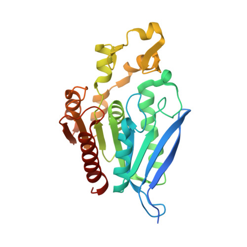
Crystal structure of the feruloyl esterase from Lentilactobacillus buchneri reveals a novel homodimeric state.
Kasmaei, K.M., Kalyani, D.C., Reichenbach, T., Jimenez-Quero, A., Vilaplana, F., Divne, C.(2022) Front Microbiol 13: 1050160-1050160
- PubMed: 36569051
- DOI: https://doi.org/10.3389/fmicb.2022.1050160
- Primary Citation of Related Structures:
7Z2U, 7Z2V, 7Z2X - PubMed Abstract:
Ferulic acid is a common constituent of the plant cell-wall matrix where it decorates and can crosslink mainly arabinoxylans to provide structural reinforcement. Microbial feruloyl esterases (FAEs) specialize in catalyzing hydrolysis of the ester bonds between phenolic acids and sugar residues in plant cell-wall polysaccharides such as arabinoxylan to release cinnamoyl compounds. Feruloyl esterases from lactic acid bacteria (LAB) have been highlighted as interesting enzymes for their potential applications in the food and pharmaceutical industries; however, there are few studies on the activity and structure of FAEs of LAB origin. Here, we report the crystal structure and biochemical characterization of a feruloyl esterase ( Lb FAE) from Lentilactobacillus buchneri , a LAB strain that has been used as a silage additive. The Lb FAE structure was determined in the absence and presence of product (FA) and reveals a new type of homodimer association not previously observed for fungal or bacterial FAEs. The two subunits associate to restrict access to the active site such that only single FA chains attached to arabinoxylan can be accommodated, an arrangement that excludes access to FA cross-links between arabinoxylan chains. This narrow specificity is further corroborated by the observation that no FA dimers are produced, only FA, when feruloylated arabinoxylan is used as substrate. Docking of arabinofuranosyl-ferulate in the Lb FAE structure highlights the restricted active site and lends further support to our hypothesis that Lb FAE is specific for single FA side chains in arabinoxylan.
Organizational Affiliation:
Department of Industrial Biotechnology, School of Engineering Sciences in Chemistry, Biotechnology, and Health (CBH), KTH Royal Institute of Technology, Stockholm, Sweden.


