7Z11 | pdb_00007z11

Structure of substrate bound DRG1 (AFG2)


Experimental Data Snapshot

  • Method: ELECTRON MICROSCOPY
  • Resolution: 3.20 Å
  • Aggregation State: PARTICLE 
  • Reconstruction Method: SINGLE PARTICLE 

wwPDB Validation 3D Report Full Report

Validation slider image for 7Z11

This is version 1.2 of the entry. See complete history

Literature

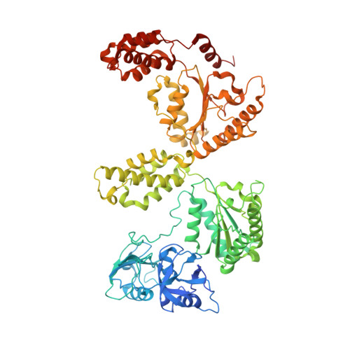
Visualizing maturation factor extraction from the nascent ribosome by the AAA-ATPase Drg1.

Prattes, M.Grishkovskaya, I.Hodirnau, V.V.Hetzmannseder, C.Zisser, G.Sailer, C.Kargas, V.Loibl, M.Gerhalter, M.Kofler, L.Warren, A.J.Stengel, F.Haselbach, D.Bergler, H.

(2022) Nat Struct Mol Biol 29: 942-953

  • DOI: https://doi.org/10.1038/s41594-022-00832-5
  • Primary Citation Related Structures: 
    7Z11, 7Z34

  • PubMed Abstract: 

    The AAA-ATPase Drg1 is a key factor in eukaryotic ribosome biogenesis that initiates cytoplasmic maturation of the large ribosomal subunit. Drg1 releases the shuttling maturation factor Rlp24 from pre-60S particles shortly after nuclear export, a strict requirement for downstream maturation. The molecular mechanism of release remained elusive. Here, we report a series of cryo-EM structures that captured the extraction of Rlp24 from pre-60S particles by Saccharomyces cerevisiae Drg1. These structures reveal that Arx1 and the eukaryote-specific rRNA expansion segment ES27 form a joint docking platform that positions Drg1 for efficient extraction of Rlp24 from the pre-ribosome. The tips of the Drg1 N domains thereby guide the Rlp24 C terminus into the central pore of the Drg1 hexamer, enabling extraction by a hand-over-hand translocation mechanism. Our results uncover substrate recognition and processing by Drg1 step by step and provide a comprehensive mechanistic picture of the conserved modus operandi of AAA-ATPases.


  • Organizational Affiliation
    • Institute of Molecular Biosciences, University of Graz, Graz, Austria.

Macromolecule Content 

  • Total Structure Weight: 516.58 kDa 
  • Atom Count: 34,079 
  • Modeled Residue Count: 4,404 
  • Deposited Residue Count: 4,700 
  • Unique protein chains: 2

Macromolecules

Find similar proteins by:|  3D Structure
Entity ID: 1
MoleculeChains  Sequence LengthOrganismDetailsImage
ATPase family gene 2 protein
A, B, C, D, E
A, B, C, D, E, F
780Saccharomyces cerevisiae S288CMutation(s): 0 
Gene Names: AFG2DRG1YLR397CL8084.16
EC: 3.6.4.10
UniProt
Find proteins for P32794 (Saccharomyces cerevisiae (strain ATCC 204508 / S288c))
Explore P32794 
Go to UniProtKB:  P32794
Entity Groups
Sequence Clusters30% Identity50% Identity70% Identity90% Identity95% Identity100% Identity
UniProt GroupP32794
Sequence Annotations
Expand
Reference Sequence
Find similar proteins by:  Sequence   |   3D Structure  
Entity ID: 2
MoleculeChains  Sequence LengthOrganismDetailsImage
peptide substrate20Saccharomyces cerevisiae S288CMutation(s): 0 

Small Molecules

Ligands 1 Unique
IDChains Name / Formula / InChI Key2D Diagram3D Interactions
AGS
(Subject of Investigation/LOI)

Query on AGS


Download:Ideal Coordinates CCD File
H [auth A]
I [auth A]
J [auth B]
K [auth B]
L [auth C]
H [auth A],
I [auth A],
J [auth B],
K [auth B],
L [auth C],
M [auth C],
N [auth D],
O [auth D],
P [auth E],
Q [auth F],
R [auth F]
PHOSPHOTHIOPHOSPHORIC ACID-ADENYLATE ESTER
C10 H16 N5 O12 P3 S
NLTUCYMLOPLUHL-KQYNXXCUSA-N

Experimental Data & Validation

Experimental Data

  • Method: ELECTRON MICROSCOPY
  • Resolution: 3.20 Å
  • Aggregation State: PARTICLE 
  • Reconstruction Method: SINGLE PARTICLE 
EM Software:
TaskSoftware PackageVersion
MODEL REFINEMENTRosetta
MODEL REFINEMENTPHENIXv1.18.2-3874

Structure Validation

View Full Validation Report


Entry History 

& Funding Information

Deposition Data


Funding OrganizationLocationGrant Number
Austrian Science FundAustria--

Revision History  (Full details and data files)

  • Version 1.0: 2022-09-21
    Type: Initial release
  • Version 1.1: 2022-10-05
    Changes: Database references
  • Version 1.2: 2024-07-17
    Changes: Data collection