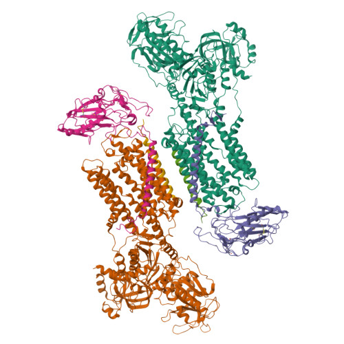
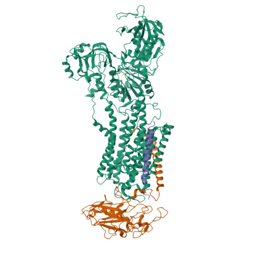
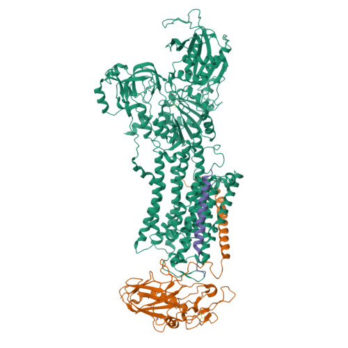
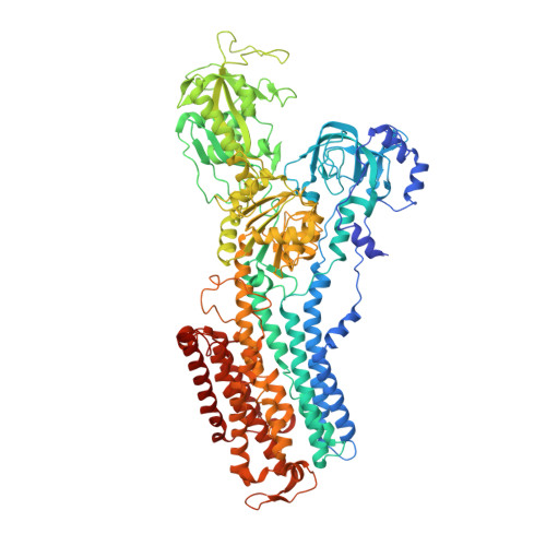
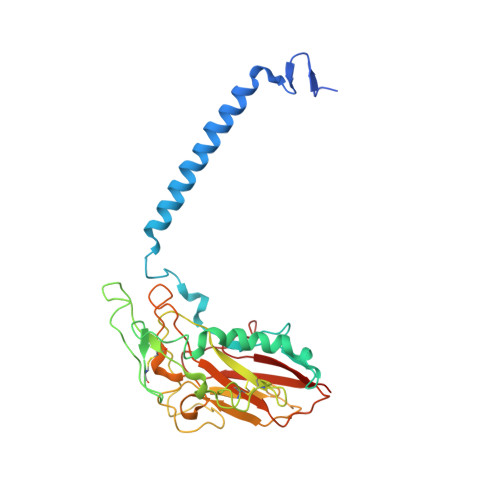
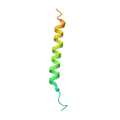
7YZR

50 mM Rb+ soak of beryllium fluoride inhibited Na+,K+-ATPase, E2-BeFx (rigid body model)


Experimental Data Snapshot

  • Method: X-RAY DIFFRACTION
  • Resolution: 6.92 Å
  • R-Value Free: 0.309 
  • R-Value Work: 0.289 
  • R-Value Observed: 0.291 

Starting Model: experimental
View more details

wwPDB Validation   3D Report Full Report


This is version 1.1 of the entry. See complete history


Literature

The Na + ,K + -ATPase in complex with beryllium fluoride mimics an ATPase phosphorylated state.

Fruergaard, M.U.Dach, I.Andersen, J.L.Ozol, M.Shahsavar, A.Quistgaard, E.M.Poulsen, H.Fedosova, N.U.Nissen, P.

(2022) J Biol Chem 298: 102317-102317

  • DOI: https://doi.org/10.1016/j.jbc.2022.102317
  • Primary Citation of Related Structures:  
    7QTV, 7YZR, 7Z04

  • PubMed Abstract: 

    The Na + ,K + -ATPase generates electrochemical gradients of Na + and K + across the plasma membrane via a functional cycle that includes various phosphoenzyme intermediates. However, the structure and function of these intermediates and how metal fluorides mimick them require further investigation. Here, we describe a 4.0 Å resolution crystal structure and functional properties of the pig kidney Na + ,K + -ATPase stabilized by the inhibitor beryllium fluoride (denoted E2-BeF x ). E2-BeF x is expected to mimic properties of the E2P phosphoenzyme, yet with unknown characteristics of ion and ligand binding. The structure resembles the E2P form obtained by phosphorylation from inorganic phosphate (P i ) and stabilized by cardiotonic steroids, including a low-affinity Mg 2+ site near ion binding site II. Our anomalous Fourier analysis of the crystals soaked in Rb + (a K + congener) followed by a low-resolution rigid-body refinement (6.9-7.5 Å) revealed preocclusion transitions leading to activation of the dephosphorylation reaction. We show that the Mg 2+ location indicates a site of initial K + recognition and acceptance upon binding to the outward-open E2P state after Na + release. Furthermore, using binding and activity studies, we find that the BeF x -inhibited enzyme is also able to bind ADP/ATP and Na + . These results relate the E2-BeF x complex to a transient K + - and ADP-sensitive E∗P intermediate of the functional cycle of the Na + ,K + -ATPase, prior to E2P.


  • Organizational Affiliation

    Department of Molecular Biology and Genetics, DANDRITE - Nordic EMBL Partnership for Molecular Medicine, Aarhus University, Aarhus C, Denmark.


Macromolecules
Find similar proteins by:  (by identity cutoff)  |  3D Structure
Entity ID: 1
MoleculeChains Sequence LengthOrganismDetailsImage
Sodium/potassium-transporting ATPase subunit alpha-1A,
D [auth C]
1,020Sus scrofaMutation(s): 0 
EC: 7.2.2.13
Membrane Entity: Yes 
UniProt
Find proteins for P05024 (Sus scrofa)
Explore P05024 
Go to UniProtKB:  P05024
Entity Groups  
Sequence Clusters30% Identity50% Identity70% Identity90% Identity95% Identity100% Identity
UniProt GroupP05024
Sequence Annotations
Expand
  • Reference Sequence
Find similar proteins by:  (by identity cutoff)  |  3D Structure
Entity ID: 2
MoleculeChains Sequence LengthOrganismDetailsImage
Sodium/potassium-transporting ATPase subunit beta-1B,
E [auth D]
302Sus scrofaMutation(s): 0 
Membrane Entity: Yes 
UniProt
Find proteins for P05027 (Sus scrofa)
Explore P05027 
Go to UniProtKB:  P05027
Entity Groups  
Sequence Clusters30% Identity50% Identity70% Identity90% Identity95% Identity100% Identity
UniProt GroupP05027
Sequence Annotations
Expand
  • Reference Sequence
Find similar proteins by:  (by identity cutoff)  |  3D Structure
Entity ID: 3
MoleculeChains Sequence LengthOrganismDetailsImage
FXYD domain-containing ion transport regulatorC [auth G],
F [auth E]
64Sus scrofaMutation(s): 0 
Membrane Entity: Yes 
UniProt
Find proteins for Q58K79 (Sus scrofa)
Explore Q58K79 
Go to UniProtKB:  Q58K79
Entity Groups  
Sequence Clusters30% Identity50% Identity70% Identity90% Identity95% Identity100% Identity
UniProt GroupQ58K79
Sequence Annotations
Expand
  • Reference Sequence
Experimental Data & Validation

Experimental Data

  • Method: X-RAY DIFFRACTION
  • Resolution: 6.92 Å
  • R-Value Free: 0.309 
  • R-Value Work: 0.289 
  • R-Value Observed: 0.291 
  • Space Group: P 21 21 21
Unit Cell:
Length ( Å )Angle ( ˚ )
a = 118.94α = 90
b = 118.92β = 90
c = 496.9γ = 90
Software Package:
Software NamePurpose
PHENIXrefinement
XSCALEdata scaling
PDB_EXTRACTdata extraction
XDSdata reduction
PHASERphasing

Structure Validation

View Full Validation Report


Entry History & Funding Information

Deposition Data


Funding OrganizationLocationGrant Number
LundbeckfondenDenmarkR155-2015-2666

Revision History  (Full details and data files)

  • Version 1.0: 2022-11-23
    Type: Initial release
  • Version 1.1: 2024-01-31
    Changes: Data collection, Refinement description