7YX8 | pdb_00007yx8

Crystal structure of the AM0627 (E326A) inactive mutant in complex with PSGL-1-like bis-T glycopeptide and Zn2+


Experimental Data Snapshot

  • Method: X-RAY DIFFRACTION
  • Resolution: 1.50 Å
  • R-Value Free: 
    0.182 (Depositor), 0.188 (DCC) 
  • R-Value Work: 
    0.167 (Depositor), 0.175 (DCC) 
  • R-Value Observed: 
    0.168 (Depositor) 

wwPDB Validation 3D Report Full Report

Validation slider image for 7YX8

This is version 1.2 of the entry. See complete history

Literature

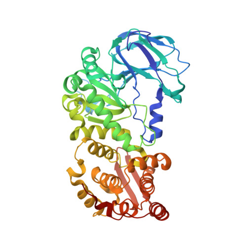
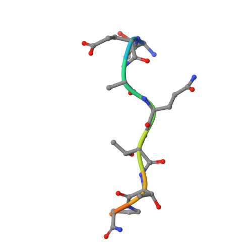
Structural and mechanistic insights into the cleavage of clustered O-glycan patches-containing glycoproteins by mucinases of the human gut.

Taleb, V.Liao, Q.Narimatsu, Y.Garcia-Garcia, A.Companon, I.Borges, R.J.Gonzalez-Ramirez, A.M.Corzana, F.Clausen, H.Rovira, C.Hurtado-Guerrero, R.

(2022) Nat Commun 13: 4324-4324

  • DOI: https://doi.org/10.1038/s41467-022-32021-9
  • Primary Citation Related Structures: 
    7YX8

  • PubMed Abstract: 

    Mucinases of human gut bacteria cleave peptide bonds in mucins strictly depending on the presence of neighboring O-glycans. The Akkermansia muciniphila AM0627 mucinase cleaves specifically in between contiguous (bis) O-glycans of defined truncated structures, suggesting that this enzyme may recognize clustered O-glycan patches. Here, we report the structure and molecular mechanism of AM0627 in complex with a glycopeptide containing a bis-T (Galβ1-3GalNAcα1-O-Ser/Thr) O-glycan, revealing that AM0627 recognizes both the sugar moieties and the peptide sequence. AM0627 exhibits preference for bis-T over bis-Tn (GalNAcα1-O-Ser/Thr) O-glycopeptide substrates, with the first GalNAc residue being essential for cleavage. AM0627 follows a mechanism relying on a nucleophilic water molecule and a catalytic base Glu residue. Structural comparison among mucinases identifies a conserved Tyr engaged in sugar-π interactions in both AM0627 and the Bacteroides thetaiotaomicron BT4244 mucinase as responsible for the common activity of these two mucinases with bis-T/Tn substrates. Our work illustrates how mucinases through tremendous flexibility adapt to the diversity in distribution and patterns of O-glycans on mucins.


  • Organizational Affiliation
    • Institute of Biocomputation and Physics of Complex Systems, University of Zaragoza, Mariano Esquillor s/n, Campus Rio Ebro, Edificio I+D, Zaragoza, Spain.

Macromolecule Content 

  • Total Structure Weight: 104.51 kDa 
  • Atom Count: 8,088 
  • Modeled Residue Count: 895 
  • Deposited Residue Count: 902 
  • Unique protein chains: 2

Macromolecules

Find similar proteins by:|  3D Structure
Entity ID: 1
MoleculeChains  Sequence LengthOrganismDetailsImage
Peptidase M60 domain-containing protein
A, B
441Akkermansia muciniphilaMutation(s): 1 
Gene Names: Amuc_0627
UniProt
Find proteins for B2UPI7 (Akkermansia muciniphila (strain ATCC BAA-835 / DSM 22959 / JCM 33894 / BCRC 81048 / CCUG 64013 / CIP 107961 / Muc))
Explore B2UPI7 
Go to UniProtKB:  B2UPI7
Entity Groups
Sequence Clusters30% Identity50% Identity70% Identity90% Identity95% Identity100% Identity
UniProt GroupB2UPI7
Sequence Annotations
Expand
Reference Sequence
Find similar proteins by:  Sequence   |   3D Structure  
Entity ID: 2
MoleculeChains  Sequence LengthOrganismDetailsImage
PSGL-1-like bis-T glycopeptideC [auth F],
D [auth H]
10synthetic constructMutation(s): 0 

Oligosaccharides

Help  
Entity ID: 3
MoleculeChains Length2D Diagram GlycosylationD Interactions
beta-D-galactopyranose-(1-3)-2-acetamido-2-deoxy-alpha-D-galactopyranoseE [auth D],
F [auth C],
G,
H [auth E]
2O-Glycosylation
Glycosylation Resources
GlyTouCan: G00031MO
GlyCosmos: G00031MO
GlyGen: G00031MO

Small Molecules

Ligands 2 Unique
IDChains Name / Formula / InChI Key2D Diagram3D Interactions
GOL

Query on GOL


Download:Ideal Coordinates CCD File
I [auth A]
J [auth A]
K [auth A]
L [auth A]
M [auth A]
I [auth A],
J [auth A],
K [auth A],
L [auth A],
M [auth A],
N [auth A],
O [auth A],
P [auth A],
R [auth B],
S [auth B]
GLYCEROL
C3 H8 O3
PEDCQBHIVMGVHV-UHFFFAOYSA-N
ZN

Query on ZN


Download:Ideal Coordinates CCD File
Q [auth A],
T [auth B]
ZINC ION
Zn
PTFCDOFLOPIGGS-UHFFFAOYSA-N

Experimental Data & Validation

Experimental Data

  • Method: X-RAY DIFFRACTION
  • Resolution: 1.50 Å
  • R-Value Free:  0.182 (Depositor), 0.188 (DCC) 
  • R-Value Work:  0.167 (Depositor), 0.175 (DCC) 
  • R-Value Observed: 0.168 (Depositor) 
Space Group: P 21 21 21
Unit Cell:
Length ( Å )Angle ( ˚ )
a = 73.145α = 90
b = 82.687β = 90
c = 172.608γ = 90
Software Package:
Software NamePurpose
REFMACrefinement
XDSdata reduction
SCALAdata scaling
SHELXDEphasing

Structure Validation

View Full Validation Report


Entry History 

& Funding Information

Deposition Data


Funding OrganizationLocationGrant Number
Spanish Ministry of Science, Innovation, and UniversitiesSpain--

Revision History  (Full details and data files)

  • Version 1.0: 2022-07-20
    Type: Initial release
  • Version 1.1: 2022-08-10
    Changes: Database references
  • Version 1.2: 2024-11-13
    Changes: Data collection, Refinement description, Structure summary