


Functional Implications of the Exon 9 Splice Insert in GluK1 Kainate Receptors
Dhingra, S., Chopade, P.M., Vinnakota, R., Kumar, J.(2023) Elife
Experimental Data Snapshot
Starting Models: experimental
View more details
wwPDB Validation 3D Report Full Report

(2023) Elife
Entity ID: 1 | |||||
|---|---|---|---|---|---|
| Molecule | Chains | Sequence Length | Organism | Details | Image |
| Glutamate receptor | 1,098 | Rattus norvegicus | Mutation(s): 2 Gene Names: Grik1, rCG_58820 | 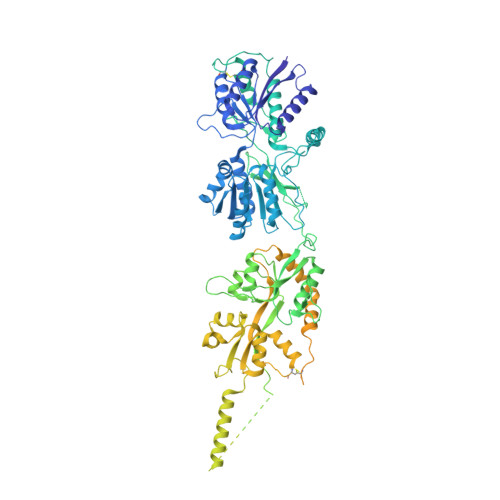 | |
UniProt | |||||
Entity Groups | |||||
| Sequence Clusters | 30% Identity50% Identity70% Identity90% Identity95% Identity100% Identity | ||||
| UniProt Group | P22756 | ||||
Sequence AnnotationsExpand | |||||
Reference Sequence | |||||
| Task | Software Package | Version |
|---|---|---|
| MODEL REFINEMENT | PHENIX |
| Funding Organization | Location | Grant Number |
|---|---|---|
| Science and Engineering Research Board (SERB) | India | CRG/2020/003971 |


