7YMP | pdb_00007ymp

Crystal structure of lysoplasmalogen specific phospholipase D


Experimental Data Snapshot

  • Method: X-RAY DIFFRACTION
  • Resolution: 2.57 Å
  • R-Value Free: 
    0.272 (Depositor), 0.272 (DCC) 
  • R-Value Work: 
    0.210 (Depositor), 0.211 (DCC) 
  • R-Value Observed: 
    0.213 (Depositor) 

Starting Model: experimental
View more details

wwPDB Validation 3D Report Full Report

Validation slider image for 7YMP

This is version 1.2 of the entry. See complete history

Literature

Structural basis for the substrate specificity switching of lysoplasmalogen-specific phospholipase D from Thermocrispum sp. RD004668.

Hamana, H.Yasutake, Y.Kato-Murayama, M.Hosaka, T.Shirouzu, M.Sakasegawa, S.I.Sugimori, D.Murayama, K.

(2022) Biosci Biotechnol Biochem 87: 74-81

  • DOI: https://doi.org/10.1093/bbb/zbac169
  • Primary Citation Related Structures: 
    7YM0, 7YMP, 7YMQ, 7YMR

  • PubMed Abstract: 

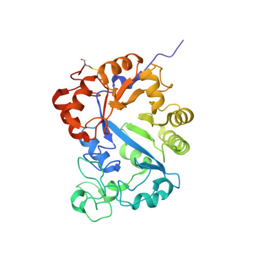
    Lysoplasmalogen-specific phospholipase D (LyPls-PLD) hydrolyzes choline lysoplasmalogen to choline and 1-(1-alkenyl)-sn-glycero-3-phosphate. Mutation of F211 to leucine altered its substrate specificity from lysoplasmalogen to 1-O-hexadecyl-2-hydroxy-sn-glycero-3-phosphocholine (lysoPAF). Enzymes specific to lysoPAF have good potential for clinical application, and understanding the mechanism of their activity is important. The crystal structure of LyPls-PLD exhibited a TIM barrel fold assigned to glycerophosphocholine phosphodiesterase, a member of glycerophosphodiester phosphodiesterase. LyPls-PLD possesses a hydrophobic cleft for the binding of the aliphatic chain of the substrate. In the structure of the F211L mutant, Met232 and Tyr258 form a "small lid" structure that stabilizes the binding of the aliphatic chain of the substrate. In contrast, F211 may inhibit small lid formation in the wild-type structure. LysoPAF possesses a flexible aliphatic chain; therefore, a small lid is effective for stabilizing the substrate during catalytic reactions.


  • Organizational Affiliation
    • Graduate School of Biomedical Engineering, Tohoku University, 2-1 Seiryo, Aoba, Sendai, Japan.

Macromolecule Content 

  • Total Structure Weight: 275.62 kDa 
  • Atom Count: 18,857 
  • Modeled Residue Count: 2,413 
  • Deposited Residue Count: 2,512 
  • Unique protein chains: 1

Macromolecules

Find similar proteins by:|  3D Structure
Entity ID: 1
MoleculeChains  Sequence LengthOrganismDetailsImage
Lysoplasmalogenase
A, B, C, D, E
A, B, C, D, E, F, G, H
314Thermocrispum sp. RD004668Mutation(s): 0 
Gene Names: lyspls
UniProt
Find proteins for A0A0U4VTN7 (Thermocrispum sp. RD004668)
Explore A0A0U4VTN7 
Go to UniProtKB:  A0A0U4VTN7
Entity Groups
Sequence Clusters30% Identity50% Identity70% Identity90% Identity95% Identity100% Identity
UniProt GroupA0A0U4VTN7
Sequence Annotations
Expand
Reference Sequence

Experimental Data & Validation

Experimental Data

  • Method: X-RAY DIFFRACTION
  • Resolution: 2.57 Å
  • R-Value Free:  0.272 (Depositor), 0.272 (DCC) 
  • R-Value Work:  0.210 (Depositor), 0.211 (DCC) 
  • R-Value Observed: 0.213 (Depositor) 
Space Group: P 1 21 1
Unit Cell:
Length ( Å )Angle ( ˚ )
a = 112.61α = 90
b = 94.94β = 90.243
c = 147.35γ = 90
Software Package:
Software NamePurpose
PHENIXrefinement
XDSdata reduction
XSCALEdata scaling
PHASERphasing

Structure Validation

View Full Validation Report


Entry History 

& Funding Information

Deposition Data


Funding OrganizationLocationGrant Number
Not funded--

Revision History  (Full details and data files)

  • Version 1.0: 2023-02-08
    Type: Initial release
  • Version 1.1: 2023-11-29
    Changes: Data collection, Refinement description
  • Version 1.2: 2024-11-06
    Changes: Structure summary