7YL3

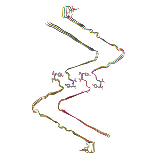
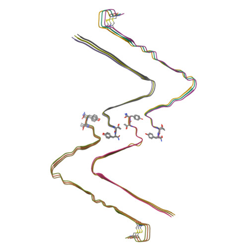
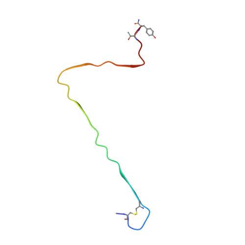
Structure of hIAPP-TF-type1


Experimental Data Snapshot

  • Method: ELECTRON MICROSCOPY
  • Resolution: 3.20 Å
  • Aggregation State: HELICAL ARRAY 
  • Reconstruction Method: HELICAL 

wwPDB Validation   3D Report Full Report


This is version 1.1 of the entry. See complete history


Literature

A new polymorphism of human amylin fibrils with similar protofilaments and a conserved core.

Li, D.Zhang, X.Wang, Y.Zhang, H.Song, K.Bao, K.Zhu, P.

(2022) iScience 25: 105705-105705

  • DOI: https://doi.org/10.1016/j.isci.2022.105705
  • Primary Citation of Related Structures:  
    7YKW, 7YL0, 7YL3, 7YL7

  • PubMed Abstract: 

    Pancreatic amyloid deposits composed of a fibrillar form of the human islet amyloid polypeptide (hIAPP) are the pathological hallmark of type 2 diabetes (T2D). Although various cryo-EM structures of polymorphic hIAPP fibrils were reported, the underlying polymorphic mechanism of hIAPP remains elusive. Meanwhile, the structure of hIAPP fibrils with all residues visible in the fibril core is not available. Here, we report the full-length structures of two different polymorphs of hIAPP fibrils, namely slim form (SF, dimer) and thick form (TF, tetramer), formed in a salt-free environment, which share a similar ζ-shaped protofilament but differ in inter-protofilament interfaces. In the absence of salt, electrostatic interactions were found to play a dominant role in stabilizing the fibril structure, suggesting an antagonistic effect between electrostatic and hydrophobic interactions in different salt concentrations environments. Our results shed light on understanding the mechanism of amyloid fibril polymorphism.


  • Organizational Affiliation

    National Laboratory of Biomacromolecules, CAS Center for Excellence in Biomacromolecules, Institute of Biophysics, Chinese Academy of Sciences, Beijing 100101, China.


Macromolecules
Find similar proteins by:  (by identity cutoff)  |  3D Structure
Entity ID: 1
MoleculeChains Sequence LengthOrganismDetailsImage
Islet amyloid polypeptide37Homo sapiensMutation(s): 0 
UniProt & NIH Common Fund Data Resources
Find proteins for P10997 (Homo sapiens)
Explore P10997 
Go to UniProtKB:  P10997
PHAROS:  P10997
GTEx:  ENSG00000121351 
Entity Groups  
Sequence Clusters30% Identity50% Identity70% Identity90% Identity95% Identity100% Identity
UniProt GroupP10997
Sequence Annotations
Expand
  • Reference Sequence
Experimental Data & Validation

Experimental Data

  • Method: ELECTRON MICROSCOPY
  • Resolution: 3.20 Å
  • Aggregation State: HELICAL ARRAY 
  • Reconstruction Method: HELICAL 
EM Software:
TaskSoftware PackageVersion
RECONSTRUCTIONRELION3.0

Structure Validation

View Full Validation Report


Entry History & Funding Information

Deposition Data


Funding OrganizationLocationGrant Number
National Natural Science Foundation of China (NSFC)China2017YFA0504700
National Natural Science Foundation of China (NSFC)China2018YFE0203300
National Natural Science Foundation of China (NSFC)China2021YFA1300100

Revision History  (Full details and data files)

  • Version 1.0: 2022-12-28
    Type: Initial release
  • Version 1.1: 2024-05-08
    Changes: Data collection, Database references