7YID

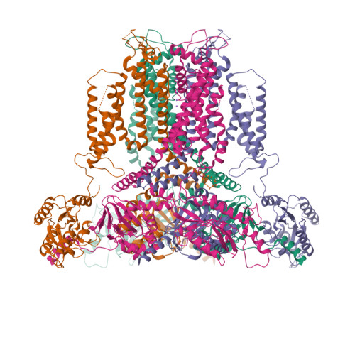
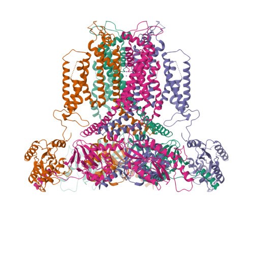
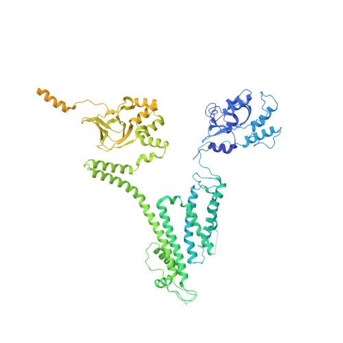
Human KCNH5 closed state 1


Experimental Data Snapshot

  • Method: ELECTRON MICROSCOPY
  • Resolution: 3.40 Å
  • Aggregation State: PARTICLE 
  • Reconstruction Method: SINGLE PARTICLE 

wwPDB Validation   3D Report Full Report


This is version 1.1 of the entry. See complete history


Literature

Mechanism underlying delayed rectifying in human voltage-mediated activation Eag2 channel.

Zhang, M.Shan, Y.Pei, D.

(2023) Nat Commun 14: 1470-1470

  • DOI: https://doi.org/10.1038/s41467-023-37204-6
  • Primary Citation of Related Structures:  
    7YID, 7YIE, 7YIF, 7YIG, 7YIH, 7YIJ

  • PubMed Abstract: 

    The transmembrane voltage gradient is a general physico-chemical cue that regulates diverse biological function through voltage-gated ion channels. How voltage sensing mediates ion flows remains unknown at the molecular level. Here, we report six conformations of the human Eag2 (hEag2) ranging from closed, pre-open, open, and pore dilation but non-conducting states captured by cryo-electron microscopy (cryo-EM). These multiple states illuminate dynamics of the selectivity filter and ion permeation pathway with delayed rectifier properties and Cole-Moore effect at the atomic level. Mechanistically, a short S4-S5 linker is coupled with the constrict sites to mediate voltage transducing in a non-domain-swapped configuration, resulting transitions for constrict sites of F464 and Q472 from gating to open state stabilizing for voltage energy transduction. Meanwhile, an additional potassium ion occupied at positions S6 confers the delayed rectifier property and Cole-Moore effects. These results provide insight into voltage transducing and potassium current across membrane, and shed light on the long-sought Cole-Moore effects.


  • Organizational Affiliation

    Fudan University, 200433, Shanghai, China.


Macromolecules
Find similar proteins by:  (by identity cutoff)  |  3D Structure
Entity ID: 1
MoleculeChains Sequence LengthOrganismDetailsImage
Potassium voltage-gated channel subfamily H member 5
A, B, C, D
988Homo sapiensMutation(s): 0 
Gene Names: KCNH5EAG2
Membrane Entity: Yes 
UniProt & NIH Common Fund Data Resources
Find proteins for Q8NCM2 (Homo sapiens)
Explore Q8NCM2 
Go to UniProtKB:  Q8NCM2
PHAROS:  Q8NCM2
GTEx:  ENSG00000140015 
Entity Groups  
Sequence Clusters30% Identity50% Identity70% Identity90% Identity95% Identity100% Identity
UniProt GroupQ8NCM2
Sequence Annotations
Expand
  • Reference Sequence
Experimental Data & Validation

Experimental Data

  • Method: ELECTRON MICROSCOPY
  • Resolution: 3.40 Å
  • Aggregation State: PARTICLE 
  • Reconstruction Method: SINGLE PARTICLE 

Structure Validation

View Full Validation Report


Entry History & Funding Information

Deposition Data

  • Released Date: 2023-04-05 
  • Deposition Author(s): Zhang, M.F.

Funding OrganizationLocationGrant Number
National Basic Research Program of China (973 Program)China2017YFA0504100

Revision History  (Full details and data files)

  • Version 1.0: 2023-04-05
    Type: Initial release
  • Version 1.1: 2024-07-03
    Changes: Data collection