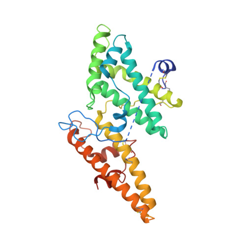
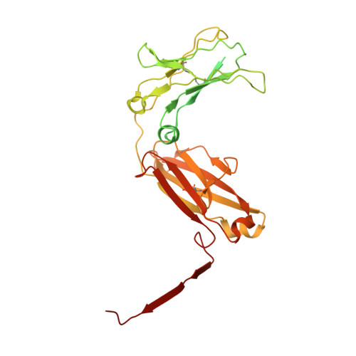
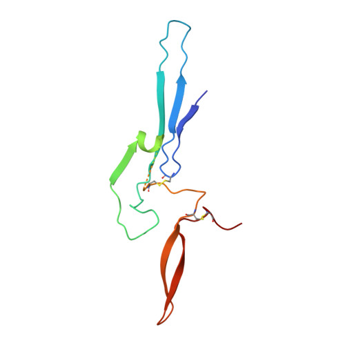
7YG2

Cryo-EM structure of human IgM-Fc in complex with the J chain and the DBL domain of DBLMSP2


Experimental Data Snapshot

  • Method: ELECTRON MICROSCOPY
  • Resolution: 3.32 Å
  • Aggregation State: PARTICLE 
  • Reconstruction Method: SINGLE PARTICLE 

wwPDB Validation   3D Report Full Report


This is version 1.3 of the entry. See complete history


Literature

Plasmodium falciparum has evolved multiple mechanisms to hijack human immunoglobulin M.

Ji, C.Shen, H.Su, C.Li, Y.Chen, S.Sharp, T.H.Xiao, J.

(2023) Nat Commun 14: 2650-2650

  • DOI: https://doi.org/10.1038/s41467-023-38320-z
  • Primary Citation of Related Structures:  
    7Y09, 7Y0H, 7Y0J, 7YG2

  • PubMed Abstract: 

    Plasmodium falciparum causes the most severe malaria in humans. Immunoglobulin M (IgM) serves as the first line of humoral defense against infection and potently activates the complement pathway to facilitate P. falciparum clearance. A number of P. falciparum proteins bind IgM, leading to immune evasion and severe disease. However, the underlying molecular mechanisms remain unknown. Here, using high-resolution cryo-electron microscopy, we delineate how P. falciparum proteins VAR2CSA, TM284VAR1, DBLMSP, and DBLMSP2 target IgM. Each protein binds IgM in a different manner, and together they present a variety of Duffy-binding-like domain-IgM interaction modes. We further show that these proteins interfere directly with IgM-mediated complement activation in vitro, with VAR2CSA exhibiting the most potent inhibitory effect. These results underscore the importance of IgM for human adaptation of P. falciparum and provide critical insights into its immune evasion mechanism.


  • Organizational Affiliation

    State Key Laboratory of Protein and Plant Gene Research, School of Life Sciences, Peking University, Beijing, China.


Macromolecules
Find similar proteins by:  (by identity cutoff)  |  3D Structure
Entity ID: 1
MoleculeChains Sequence LengthOrganismDetailsImage
DBLMSP2A [auth M]307Plasmodium falciparumMutation(s): 0 
Gene Names: DBLMSP2
UniProt
Find proteins for A0A0A7MCY3 (Plasmodium falciparum)
Explore A0A0A7MCY3 
Go to UniProtKB:  A0A0A7MCY3
Entity Groups  
Sequence Clusters30% Identity50% Identity70% Identity90% Identity95% Identity100% Identity
UniProt GroupA0A0A7MCY3
Sequence Annotations
Expand
  • Reference Sequence
Find similar proteins by:  (by identity cutoff)  |  3D Structure
Entity ID: 2
MoleculeChains Sequence LengthOrganismDetailsImage
Immunoglobulin heavy constant mu383Homo sapiensMutation(s): 0 
Gene Names: IGHM
UniProt & NIH Common Fund Data Resources
Find proteins for P01871 (Homo sapiens)
Explore P01871 
Go to UniProtKB:  P01871
PHAROS:  P01871
GTEx:  ENSG00000211899 
Entity Groups  
Sequence Clusters30% Identity50% Identity70% Identity90% Identity95% Identity100% Identity
UniProt GroupP01871
Glycosylation
Glycosylation Sites: 1Go to GlyGen: P01871-2
Sequence Annotations
Expand
  • Reference Sequence
Find similar proteins by:  (by identity cutoff)  |  3D Structure
Entity ID: 3
MoleculeChains Sequence LengthOrganismDetailsImage
Immunoglobulin J chainL [auth J]136Homo sapiensMutation(s): 0 
Gene Names: JCHAINIGCJIGJ
UniProt & NIH Common Fund Data Resources
Find proteins for P01591 (Homo sapiens)
Explore P01591 
Go to UniProtKB:  P01591
PHAROS:  P01591
GTEx:  ENSG00000132465 
Entity Groups  
Sequence Clusters30% Identity50% Identity70% Identity90% Identity95% Identity100% Identity
UniProt GroupP01591
Glycosylation
Glycosylation Sites: 1Go to GlyGen: P01591-1
Sequence Annotations
Expand
  • Reference Sequence
Oligosaccharides

Help

Entity ID: 4
MoleculeChains Length2D Diagram Glycosylation3D Interactions
2-acetamido-2-deoxy-beta-D-glucopyranose-(1-4)-2-acetamido-2-deoxy-beta-D-glucopyranoseM [auth I]2N-Glycosylation
Glycosylation Resources
GlyTouCan:  G42666HT
GlyCosmos:  G42666HT
GlyGen:  G42666HT
Small Molecules
Ligands 1 Unique
IDChains Name / Formula / InChI Key2D Diagram3D Interactions
NAG (Subject of Investigation/LOI)
Query on NAG

Download Ideal Coordinates CCD File 
N [auth A]
O [auth B]
P [auth C]
Q [auth D]
R [auth E]
N [auth A],
O [auth B],
P [auth C],
Q [auth D],
R [auth E],
S [auth F],
T [auth G],
U [auth H],
V [auth K],
W [auth L]
2-acetamido-2-deoxy-beta-D-glucopyranose
C8 H15 N O6
OVRNDRQMDRJTHS-FMDGEEDCSA-N
Experimental Data & Validation

Experimental Data

  • Method: ELECTRON MICROSCOPY
  • Resolution: 3.32 Å
  • Aggregation State: PARTICLE 
  • Reconstruction Method: SINGLE PARTICLE 

Structure Validation

View Full Validation Report


Entry History & Funding Information

Deposition Data


Funding OrganizationLocationGrant Number
National Science Foundation (NSF, China)China--

Revision History  (Full details and data files)

  • Version 1.0: 2023-03-29
    Type: Initial release
  • Version 1.1: 2023-10-11
    Changes: Data collection, Database references
  • Version 1.2: 2024-05-08
    Changes: Database references
  • Version 1.3: 2024-10-09
    Changes: Data collection, Structure summary