7Y1T | pdb_00007y1t

Complex of integrin alphaV/beta8 and L-TGF-beta1 at a ratio of 1:2


Experimental Data Snapshot

  • Method: ELECTRON MICROSCOPY
  • Resolution: 3.24 Å
  • Aggregation State: PARTICLE 
  • Reconstruction Method: SINGLE PARTICLE 

wwPDB Validation 3D Report Full Report

Validation slider image for 7Y1T

This is version 1.2 of the entry. See complete history

Literature

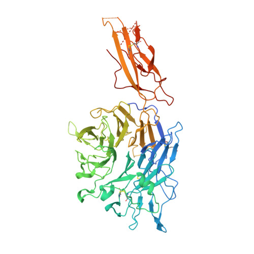
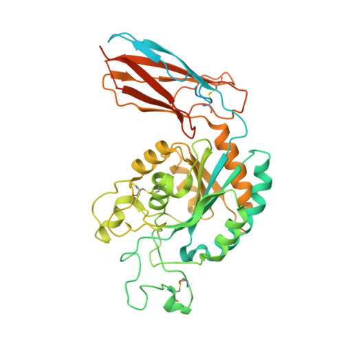
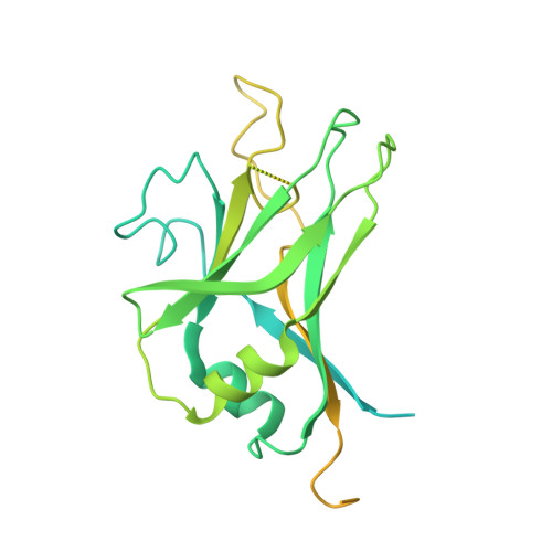
Specificity of TGF-beta 1 signal designated by LRRC33 and integrin alpha V beta 8.

Duan, Z.Lin, X.Wang, L.Zhen, Q.Jiang, Y.Chen, C.Yang, J.Lee, C.H.Qin, Y.Li, Y.Zhao, B.Wang, J.Zhang, Z.

(2022) Nat Commun 13: 4988-4988

  • DOI: https://doi.org/10.1038/s41467-022-32655-9
  • Primary Citation Related Structures: 
    7Y1R, 7Y1T

  • PubMed Abstract: 

    Myeloid lineage cells present the latent form of transforming growth factor-β1 (L-TGF-β1) to the membrane using an anchor protein LRRC33. Integrin α V β 8 activates extracellular L-TGF-β1 to trigger the downstream signaling functions. However, the mechanism designating the specificity of TGF-β1 presentation and activation remains incompletely understood. Here, we report cryo-EM structures of human L-TGF-β1/LRRC33 and integrin α V β 8 /L-TGF-β1 complexes. Combined with biochemical and cell-based analyses, we demonstrate that LRRC33 only presents L-TGF-β1 but not the -β2 or -β3 isoforms due to difference of key residues on the growth factor domains. Moreover, we reveal a 2:2 binding mode of integrin α V β 8 and L-TGF-β1, which shows higher avidity and more efficient L-TGF-β1 activation than previously reported 1:2 binding mode. We also uncover that the disulfide-linked loop of the integrin subunit β 8 determines its exquisite affinity to L-TGF-β1. Together, our findings provide important insights into the specificity of TGF-β1 signaling achieved by LRRC33 and integrin α V β 8 .


  • Organizational Affiliation
    • State Key Laboratory of Membrane Biology, Center for Life Sciences, School of Life Sciences, Peking University, 100871, Beijing, China.

Macromolecule Content 

  • Total Structure Weight: 169.75 kDa 
  • Atom Count: 8,112 
  • Modeled Residue Count: 1,030 
  • Deposited Residue Count: 1,489 
  • Unique protein chains: 3

Macromolecules

Find similar proteins by:|  3D Structure
Entity ID: 1
MoleculeChains  Sequence LengthOrganismDetailsImage
Integrin alpha-V635Homo sapiensMutation(s): 1 
Gene Names: ITGAV
UniProt & NIH Common Fund Data Resources
Find proteins for P06756 (Homo sapiens)
Explore P06756 
Go to UniProtKB:  P06756
PHAROS:  P06756
GTEx:  ENSG00000138448 
Entity Groups
Sequence Clusters30% Identity50% Identity70% Identity90% Identity95% Identity100% Identity
UniProt GroupP06756
Glycosylation
Glycosylation Sites: 4Go to GlyGen: P06756-1
Sequence Annotations
Expand
Reference Sequence
Find similar proteins by:|  3D Structure
Entity ID: 2
MoleculeChains  Sequence LengthOrganismDetailsImage
Integrin beta-8477Homo sapiensMutation(s): 1 
Gene Names: ITGB8
UniProt & NIH Common Fund Data Resources
Find proteins for P26012 (Homo sapiens)
Explore P26012 
Go to UniProtKB:  P26012
PHAROS:  P26012
GTEx:  ENSG00000105855 
Entity Groups
Sequence Clusters30% Identity50% Identity70% Identity90% Identity95% Identity100% Identity
UniProt GroupP26012
Glycosylation
Glycosylation Sites: 4Go to GlyGen: P26012-1
Sequence Annotations
Expand
Reference Sequence
Find similar proteins by:|  3D Structure
Entity ID: 3
MoleculeChains  Sequence LengthOrganismDetailsImage
Transforming growth factor beta-1 proproteinC [auth D]377Homo sapiensMutation(s): 0 
Gene Names: TGFB1TGFB
UniProt & NIH Common Fund Data Resources
Find proteins for P01137 (Homo sapiens)
Explore P01137 
Go to UniProtKB:  P01137
PHAROS:  P01137
GTEx:  ENSG00000105329 
Entity Groups
Sequence Clusters30% Identity50% Identity70% Identity90% Identity95% Identity100% Identity
UniProt GroupP01137
Glycosylation
Glycosylation Sites: 1Go to GlyGen: P01137-1
Sequence Annotations
Expand
Reference Sequence

Oligosaccharides

Help  
Entity ID: 4
MoleculeChains Length2D Diagram GlycosylationD Interactions
2-acetamido-2-deoxy-beta-D-glucopyranose-(1-4)-2-acetamido-2-deoxy-beta-D-glucopyranose-(1-4)-2-acetamido-2-deoxy-beta-D-glucopyranoseD [auth F]3N-Glycosylation
Glycosylation Resources
GlyTouCan: G47362BJ
GlyCosmos: G47362BJ
GlyGen: G47362BJ
Entity ID: 5
MoleculeChains Length2D Diagram GlycosylationD Interactions
2-acetamido-2-deoxy-beta-D-glucopyranose-(1-4)-2-acetamido-2-deoxy-beta-D-glucopyranoseE [auth C],
F [auth E]
2N-Glycosylation
Glycosylation Resources
GlyTouCan: G42666HT
GlyCosmos: G42666HT
GlyGen: G42666HT
Entity ID: 6
MoleculeChains Length2D Diagram GlycosylationD Interactions
alpha-D-mannopyranose-(1-6)-alpha-D-mannopyranose-(1-4)-[alpha-D-mannopyranose-(1-3)]beta-D-mannopyranose-(1-4)-2-acetamido-2-deoxy-beta-D-glucopyranose-(1-4)-2-acetamido-2-deoxy-beta-D-glucopyranose
G
6N-Glycosylation

Small Molecules

Ligands 3 Unique
IDChains Name / Formula / InChI Key2D Diagram3D Interactions
NAG

Query on NAG


Download:Ideal Coordinates CCD File
H [auth A],
M [auth B],
N [auth B],
O [auth B],
R [auth D]
2-acetamido-2-deoxy-beta-D-glucopyranose
C8 H15 N O6
OVRNDRQMDRJTHS-FMDGEEDCSA-N
MN

Query on MN


Download:Ideal Coordinates CCD File
Q [auth B]MANGANESE (II) ION
Mn
WAEMQWOKJMHJLA-UHFFFAOYSA-N
CA
(Subject of Investigation/LOI)

Query on CA


Download:Ideal Coordinates CCD File
I [auth A],
J [auth A],
K [auth A],
L [auth A],
P [auth B]
CALCIUM ION
Ca
BHPQYMZQTOCNFJ-UHFFFAOYSA-N

Experimental Data & Validation

Experimental Data

  • Method: ELECTRON MICROSCOPY
  • Resolution: 3.24 Å
  • Aggregation State: PARTICLE 
  • Reconstruction Method: SINGLE PARTICLE 

Structure Validation

View Full Validation Report


Entry History 

& Funding Information

Deposition Data


Funding OrganizationLocationGrant Number
National Natural Science Foundation of China (NSFC)China32171201

Revision History  (Full details and data files)

  • Version 1.0: 2022-08-31
    Type: Initial release
  • Version 1.1: 2022-09-07
    Changes: Database references
  • Version 1.2: 2024-11-13
    Changes: Data collection, Structure summary