7XYQ | pdb_00007xyq

Crystal strucutre of PD-L1 and the computationally designed DBL1_03 protein binder


Experimental Data Snapshot

  • Method: X-RAY DIFFRACTION
  • Resolution: 2.85 Å
  • R-Value Free: 
    0.318 (Depositor), 0.304 (DCC) 
  • R-Value Work: 
    0.300 (Depositor) 
  • R-Value Observed: 
    0.301 (Depositor) 

Starting Model: experimental
View more details

wwPDB Validation 3D Report Full Report

Validation slider image for 7XYQ

Ligand Structure Quality Assessment 


This is version 1.4 of the entry. See complete history

Literature

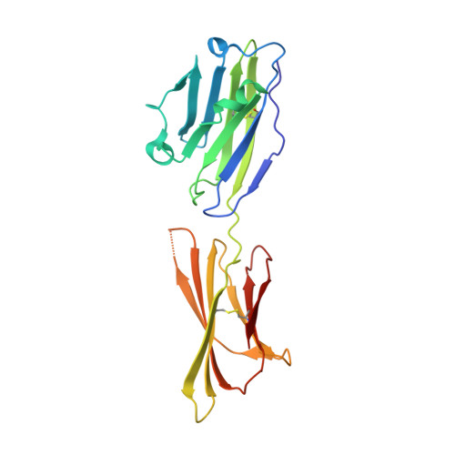
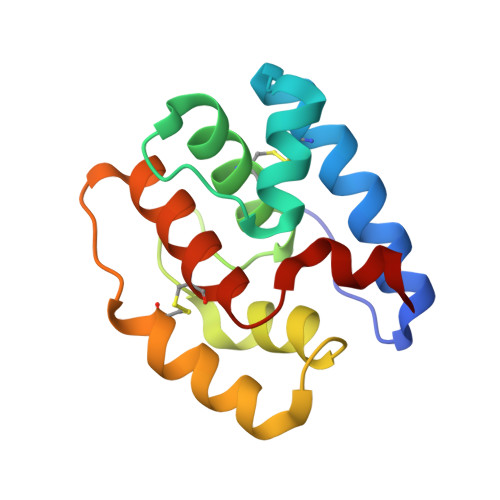
De novo design of protein interactions with learned surface fingerprints.

Gainza, P.Wehrle, S.Van Hall-Beauvais, A.Marchand, A.Scheck, A.Harteveld, Z.Buckley, S.Ni, D.Tan, S.Sverrisson, F.Goverde, C.Turelli, P.Raclot, C.Teslenko, A.Pacesa, M.Rosset, S.Georgeon, S.Marsden, J.Petruzzella, A.Liu, K.Xu, Z.Chai, Y.Han, P.Gao, G.F.Oricchio, E.Fierz, B.Trono, D.Stahlberg, H.Bronstein, M.Correia, B.E.

(2023) Nature 617: 176-184

  • DOI: https://doi.org/10.1038/s41586-023-05993-x
  • Primary Citation Related Structures: 
    7XAD, 7XYQ, 7ZRV, 7ZSD, 7ZSS

  • PubMed Abstract: 

    Physical interactions between proteins are essential for most biological processes governing life 1 . However, the molecular determinants of such interactions have been challenging to understand, even as genomic, proteomic and structural data increase. This knowledge gap has been a major obstacle for the comprehensive understanding of cellular protein-protein interaction networks and for the de novo design of protein binders that are crucial for synthetic biology and translational applications 2-9 . Here we use a geometric deep-learning framework operating on protein surfaces that generates fingerprints to describe geometric and chemical features that are critical to drive protein-protein interactions 10 . We hypothesized that these fingerprints capture the key aspects of molecular recognition that represent a new paradigm in the computational design of novel protein interactions. As a proof of principle, we computationally designed several de novo protein binders to engage four protein targets: SARS-CoV-2 spike, PD-1, PD-L1 and CTLA-4. Several designs were experimentally optimized, whereas others were generated purely in silico, reaching nanomolar affinity with structural and mutational characterization showing highly accurate predictions. Overall, our surface-centric approach captures the physical and chemical determinants of molecular recognition, enabling an approach for the de novo design of protein interactions and, more broadly, of artificial proteins with function.


  • Organizational Affiliation
    • Laboratory of Protein Design and Immunoengineering, Institute of Bioengineering, École Polytechnique Fédérale de Lausanne, Lausanne, Switzerland.

Macromolecule Content 

  • Total Structure Weight: 37.93 kDa 
  • Atom Count: 2,620 
  • Modeled Residue Count: 327 
  • Deposited Residue Count: 331 
  • Unique protein chains: 2

Macromolecules

Find similar proteins by:|  3D Structure
Entity ID: 1
MoleculeChains  Sequence LengthOrganismDetailsImage
CD274 molecule207Homo sapiensMutation(s): 0 
Gene Names: CD274
UniProt & NIH Common Fund Data Resources
Find proteins for Q9NZQ7 (Homo sapiens)
Explore Q9NZQ7 
Go to UniProtKB:  Q9NZQ7
PHAROS:  Q9NZQ7
GTEx:  ENSG00000120217 
Entity Groups
Sequence Clusters30% Identity50% Identity70% Identity90% Identity95% Identity100% Identity
UniProt GroupQ9NZQ7
Sequence Annotations
Expand
Reference Sequence
Find similar proteins by:|  3D Structure
Entity ID: 2
MoleculeChains  Sequence LengthOrganismDetailsImage
DBL1_03124synthetic constructMutation(s): 0 

Small Molecules

Ligands 1 Unique
IDChains Name / Formula / InChI Key2D Diagram3D Interactions
ARG

Query on ARG


Download:Ideal Coordinates CCD File
C [auth A]ARGININE
C6 H15 N4 O2
ODKSFYDXXFIFQN-BYPYZUCNSA-O

Experimental Data & Validation

Experimental Data

  • Method: X-RAY DIFFRACTION
  • Resolution: 2.85 Å
  • R-Value Free:  0.318 (Depositor), 0.304 (DCC) 
  • R-Value Work:  0.300 (Depositor) 
  • R-Value Observed: 0.301 (Depositor) 
Space Group: P 42 21 2
Unit Cell:
Length ( Å )Angle ( ˚ )
a = 97.93α = 90
b = 97.93β = 90
c = 106.11γ = 90
Software Package:
Software NamePurpose
PHENIXrefinement
XDSdata reduction
XSCALEdata scaling
PHASERphasing

Structure Validation

View Full Validation Report


Ligand Structure Quality Assessment 


Entry History 

& Funding Information

Deposition Data


Funding OrganizationLocationGrant Number
National Natural Science Foundation of China (NSFC)China92169208

Revision History  (Full details and data files)

  • Version 1.0: 2023-04-12
    Type: Initial release
  • Version 1.1: 2023-05-03
    Changes: Database references
  • Version 1.2: 2023-05-10
    Changes: Database references
  • Version 1.3: 2023-05-17
    Changes: Database references
  • Version 1.4: 2024-11-20
    Changes: Data collection, Structure summary