7XX3 | pdb_00007xx3

Crystal structure of human Superoxide Dismutase (SOD1) in complex with a fungal metabolite molecule, Phialomustin B (PB)


Experimental Data Snapshot

  • Method: X-RAY DIFFRACTION
  • Resolution: 1.90 Å
  • R-Value Free: 
    0.230 (Depositor), 0.215 (DCC) 
  • R-Value Work: 
    0.199 (Depositor) 
  • R-Value Observed: 
    0.200 (Depositor) 

Starting Model: experimental
View more details

wwPDB Validation 3D Report Full Report

Validation slider image for 7XX3

Ligand Structure Quality Assessment 


This is version 1.3 of the entry. See complete history

Literature

Structural insights into the modulation Of SOD1 aggregation By a fungal metabolite Phialomustin-B: Therapeutic potential in ALS.

Unni, S.Kommu, P.Aouti, S.Nalli, Y.Bharath, M.M.S.Ali, A.Padmanabhan, B.

(2024) PLoS One 19: e0298196-e0298196

  • DOI: https://doi.org/10.1371/journal.pone.0298196
  • Primary Citation Related Structures: 
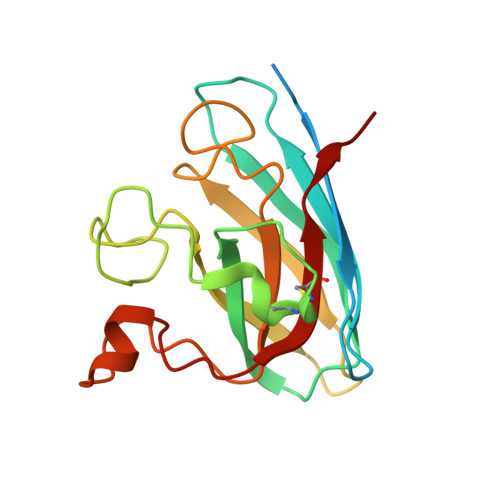
    7XX3

  • PubMed Abstract: 

    Amyotrophic lateral sclerosis (ALS) is a fatal human motor neuron disease leading to muscle atrophy and paralysis. Mutations in superoxide dismutase 1 (SOD1) are associated with familial ALS (fALS). The SOD1 mutants in ALS have a toxic-gain of function by destabilizing the functional SOD1 homodimer, consequently inducing fibril-like aggregation with a cytotoxic non-native trimer intermediate. Therefore, reducing SOD1 oligomerization via chemical modulators is an optimal therapy in ALS. Here, we report the discovery of Phialomustin-B, an unsaturated secondary metabolite from the endophytic fungus Phialophora mustea, as a modulator of SOD1 aggregation. The crystal structure of the SOD1-Phialomustin complex refined to 1.90 Å resolution demonstrated for the first time that the ligand binds to the dimer interface and the lateral region near the electrostatic loop. The aggregation analyses of SOD1WT and the disease mutant SOD1A4V revealed that Phialomustin-B reduces cytotoxic trimerization. We propose that Phialomustin-B is a potent lead molecule with therapeutic potential in fALS.


  • Organizational Affiliation
    • Department of Biophysics, National Institute of Mental Health and Neurosciences, Bengaluru, India.

Macromolecule Content 

  • Total Structure Weight: 192.83 kDa 
  • Atom Count: 12,385 
  • Modeled Residue Count: 1,506 
  • Deposited Residue Count: 1,800 
  • Unique protein chains: 1

Macromolecules

Find similar proteins by:|  3D Structure
Entity ID: 1
MoleculeChains  Sequence LengthOrganismDetailsImage
Superoxide dismutase [Cu-Zn]180Homo sapiensMutation(s): 0 
Gene Names: SOD1
EC: 1.15.1.1 (PDB Primary Data), 1.8 (UniProt)
UniProt & NIH Common Fund Data Resources
Find proteins for P00441 (Homo sapiens)
Explore P00441 
Go to UniProtKB:  P00441
PHAROS:  P00441
GTEx:  ENSG00000142168 
Entity Groups
Sequence Clusters30% Identity50% Identity70% Identity90% Identity95% Identity100% Identity
UniProt GroupP00441
Sequence Annotations
Expand
Reference Sequence

Small Molecules

Ligands 4 Unique
IDChains Name / Formula / InChI Key2D Diagram3D Interactions
I2G
(Subject of Investigation/LOI)

Query on I2G


Download:Ideal Coordinates CCD File
CA [auth I],
N [auth B],
S [auth D],
V [auth C]
(2~{E},4~{E},6~{S})-4,6-dimethyldeca-2,4-dienoic acid
C12 H20 O2
DZXNZBWYVLLUAX-IVONAZQTSA-N
GOL

Query on GOL


Download:Ideal Coordinates CCD File
L [auth A],
O [auth B],
P [auth B],
Q [auth B],
T [auth D]
GLYCEROL
C3 H8 O3
PEDCQBHIVMGVHV-UHFFFAOYSA-N
DMS

Query on DMS


Download:Ideal Coordinates CCD File
X [auth E]DIMETHYL SULFOXIDE
C2 H6 O S
IAZDPXIOMUYVGZ-UHFFFAOYSA-N
ZN

Query on ZN


Download:Ideal Coordinates CCD File
AA [auth G]
BA [auth I]
K [auth A]
M [auth B]
R [auth D]
AA [auth G],
BA [auth I],
K [auth A],
M [auth B],
R [auth D],
U [auth C],
W [auth H],
Y [auth E],
Z [auth F]
ZINC ION
Zn
PTFCDOFLOPIGGS-UHFFFAOYSA-N

Experimental Data & Validation

Experimental Data

  • Method: X-RAY DIFFRACTION
  • Resolution: 1.90 Å
  • R-Value Free:  0.230 (Depositor), 0.215 (DCC) 
  • R-Value Work:  0.199 (Depositor) 
  • R-Value Observed: 0.200 (Depositor) 
Space Group: P 63 2 2
Unit Cell:
Length ( Å )Angle ( ˚ )
a = 242.445α = 90
b = 242.445β = 90
c = 144.491γ = 120
Software Package:
Software NamePurpose
PHENIXrefinement
HKL-2000data scaling
PDB_EXTRACTdata extraction
HKL-3000data reduction
PHENIXphasing

Structure Validation

View Full Validation Report


Ligand Structure Quality Assessment 


Entry History 

& Funding Information

Deposition Data


Funding OrganizationLocationGrant Number
Indian Council of Medical ResearchIndiaIndian Council of Medical Research (ICMR),
Department of Science & Technology (DST, India)IndiaDST-FIST: SR/FST/LS-I/2017(C)

Revision History  (Full details and data files)

  • Version 1.0: 2023-12-13
    Type: Initial release
  • Version 1.1: 2024-10-30
    Changes: Structure summary
  • Version 1.2: 2025-02-26
    Changes: Database references
  • Version 1.3: 2026-03-04
    Changes: Refinement description