7XUF | pdb_00007xuf

Cryo-EM structure of the AKT1-AtKC1 complex from Arabidopsis thaliana


Experimental Data Snapshot

  • Method: ELECTRON MICROSCOPY
  • Resolution: 3.30 Å
  • Aggregation State: PARTICLE 
  • Reconstruction Method: SINGLE PARTICLE 

wwPDB Validation 3D Report Full Report

Validation slider image for 7XUF

This is version 1.2 of the entry. See complete history

Literature

Structural basis for the activity regulation of a potassium channel AKT1 from Arabidopsis.

Lu, Y.Yu, M.Jia, Y.Yang, F.Zhang, Y.Xu, X.Li, X.Yang, F.Lei, J.Wang, Y.Yang, G.

(2022) Nat Commun 13: 5682-5682

  • DOI: https://doi.org/10.1038/s41467-022-33420-8
  • Primary Citation Related Structures: 
    7FCV, 7WSW, 7XUF

  • PubMed Abstract: 

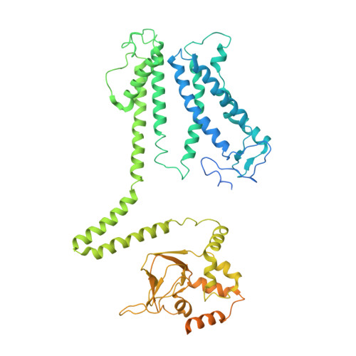
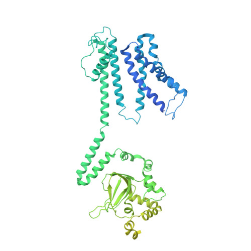
    The voltage-gated potassium channel AKT1 is responsible for primary K + uptake in Arabidopsis roots. AKT1 is functionally activated through phosphorylation and negatively regulated by a potassium channel α-subunit AtKC1. However, the molecular basis for the modulation mechanism remains unclear. Here we report the structures of AKT1, phosphorylated-AKT1, a constitutively-active variant, and AKT1-AtKC1 complex. AKT1 is assembled in 2-fold symmetry at the cytoplasmic domain. Such organization appears to sterically hinder the reorientation of C-linkers during ion permeation. Phosphorylated-AKT1 adopts an alternate 4-fold symmetric conformation at cytoplasmic domain, which indicates conformational changes associated with symmetry switch during channel activation. To corroborate this finding, we perform structure-guided mutagenesis to disrupt the dimeric interface and identify a constitutively-active variant Asp379Ala mediates K + permeation independently of phosphorylation. This variant predominantly adopts a 4-fold symmetric conformation. Furthermore, the AKT1-AtKC1 complex assembles in 2-fold symmetry. Together, our work reveals structural insight into the regulatory mechanism for AKT1.


  • Organizational Affiliation
    • State Key Laboratory for Agrobiotechnology, Frontiers Science Center for Molecular Design Breeding, College of Biological Sciences, China Agricultural University, Beijing, China.

Macromolecule Content 

  • Total Structure Weight: 345.71 kDa 
  • Atom Count: 15,041 
  • Modeled Residue Count: 1,864 
  • Deposited Residue Count: 3,038 
  • Unique protein chains: 2

Macromolecules

Find similar proteins by:|  3D Structure
Entity ID: 1
MoleculeChains  Sequence LengthOrganismDetailsImage
Potassium channel KAT3
A, C
662Arabidopsis thalianaMutation(s): 0 
Gene Names: KAT3AKT4KC1At4g32650F4D11.150
Membrane Entity: Yes 
UniProt
Find proteins for P92960 (Arabidopsis thaliana)
Explore P92960 
Go to UniProtKB:  P92960
Entity Groups
Sequence Clusters30% Identity50% Identity70% Identity90% Identity95% Identity100% Identity
UniProt GroupP92960
Sequence Annotations
Expand
Reference Sequence
Find similar proteins by:|  3D Structure
Entity ID: 2
MoleculeChains  Sequence LengthOrganismDetailsImage
Potassium channel AKT1
B, D
857Arabidopsis thalianaMutation(s): 0 
Gene Names: AKT1
Membrane Entity: Yes 
UniProt
Find proteins for Q38998 (Arabidopsis thaliana)
Explore Q38998 
Go to UniProtKB:  Q38998
Entity Groups
Sequence Clusters30% Identity50% Identity70% Identity90% Identity95% Identity100% Identity
UniProt GroupQ38998
Sequence Annotations
Expand
Reference Sequence

Experimental Data & Validation

Experimental Data

  • Method: ELECTRON MICROSCOPY
  • Resolution: 3.30 Å
  • Aggregation State: PARTICLE 
  • Reconstruction Method: SINGLE PARTICLE 
EM Software:
TaskSoftware PackageVersion
MODEL REFINEMENTPHENIX

Structure Validation

View Full Validation Report


Entry History 

& Funding Information

Deposition Data


Funding OrganizationLocationGrant Number
Not funded--

Revision History  (Full details and data files)

  • Version 1.0: 2022-11-09
    Type: Initial release
  • Version 1.1: 2024-07-03
    Changes: Data collection
  • Version 1.2: 2025-06-18
    Changes: Data collection, Structure summary