7XUD | pdb_00007xud

Structure of G9a in complex with compound 26a


Experimental Data Snapshot

  • Method: X-RAY DIFFRACTION
  • Resolution: 1.45 Å
  • R-Value Free: 
    0.177 (Depositor), 0.174 (DCC) 
  • R-Value Work: 
    0.161 (Depositor), 0.158 (DCC) 
  • R-Value Observed: 
    0.162 (Depositor) 

Starting Model: experimental
View more details

wwPDB Validation 3D Report Full Report

Validation slider image for 7XUD

Ligand Structure Quality Assessment 


This is version 1.2 of the entry. See complete history

Literature

Discovery of Novel Substrate-Competitive Lysine Methyltransferase G9a Inhibitors as Anticancer Agents.

Nishigaya, Y.Takase, S.Sumiya, T.Kikuzato, K.Sato, T.Niwa, H.Sato, S.Nakata, A.Sonoda, T.Hashimoto, N.Namie, R.Honma, T.Umehara, T.Shirouzu, M.Koyama, H.Yoshida, M.Ito, A.Shirai, F.

(2023) J Med Chem 66: 4059-4085

  • DOI: https://doi.org/10.1021/acs.jmedchem.2c02059
  • Primary Citation Related Structures: 
    7XUA, 7XUB, 7XUC, 7XUD

  • PubMed Abstract: 

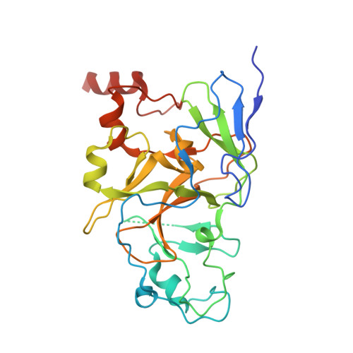
    Identification of structurally novel inhibitors of lysine methyltransferase G9a has been a subject of intense research in cancer epigenetics. Starting with the high-throughput screening (HTS) hit rac - 10a obtained from the chemical library of the University of Tokyo Drug Discovery Initiative, the structure-activity relationship of the unique substrate-competitive inhibitors was established with the help of X-ray crystallography and fragment molecular orbital (FMO) calculations for the ligand-protein interaction. Further optimization of the in vitro characteristics and drug metabolism and pharmacokinetics (DMPK) properties led to the identification of 26j (RK-701), which is a structurally distinct potent inhibitor of G9a/GLP (IC 50 = 27/53 nM). Compound 26j exhibited remarkable selectivity against other related methyltransferases, dose-dependent attenuation of cellular H3K9me2 levels, and tumor growth inhibition in MOLT-4 cells in vitro . Moreover, compound 26j showed inhibition of tumor initiation and growth in a carcinogen-induced hepatocellular carcinoma (HCC) in vivo mouse model without overt acute toxicity.


  • Organizational Affiliation
    • Watarase Research Center, Discovery Research Headquarters, Kyorin Pharmaceutical Co. Ltd., 1848 Nogi, Shimotsuga-gun, Tochigi 329-0114, Japan.

Macromolecule Content 

  • Total Structure Weight: 67.68 kDa 
  • Atom Count: 5,120 
  • Modeled Residue Count: 538 
  • Deposited Residue Count: 566 
  • Unique protein chains: 1

Macromolecules

Find similar proteins by:|  3D Structure
Entity ID: 1
MoleculeChains  Sequence LengthOrganismDetailsImage
Histone-lysine N-methyltransferase EHMT2
A, B
283Homo sapiensMutation(s): 0 
Gene Names: EHMT2BAT8C6orf30G9AKMT1CNG36
EC: 2.1.1 (PDB Primary Data), 2.1.1.367 (UniProt)
UniProt & NIH Common Fund Data Resources
Find proteins for Q96KQ7 (Homo sapiens)
Explore Q96KQ7 
Go to UniProtKB:  Q96KQ7
PHAROS:  Q96KQ7
GTEx:  ENSG00000204371 
Entity Groups
Sequence Clusters30% Identity50% Identity70% Identity90% Identity95% Identity100% Identity
UniProt GroupQ96KQ7
Sequence Annotations
Expand
Reference Sequence

Small Molecules

Ligands 6 Unique
IDChains Name / Formula / InChI Key2D Diagram3D Interactions
I6Z
(Subject of Investigation/LOI)

Query on I6Z


Download:Ideal Coordinates CCD File
I [auth A],
P [auth B]
~{N}-[(2~{S})-4-cyclopropyl-1-oxidanylidene-1-phenylazanyl-butan-2-yl]-3,6,6-trimethyl-4-oxidanylidene-5,7-dihydro-1~{H}-indole-2-carboxamide
C25 H31 N3 O3
FSDGOJSCOXFEFN-SFHVURJKSA-N
SFG

Query on SFG


Download:Ideal Coordinates CCD File
H [auth A],
O [auth B]
SINEFUNGIN
C15 H23 N7 O5
LMXOHSDXUQEUSF-YECHIGJVSA-N
TRS

Query on TRS


Download:Ideal Coordinates CCD File
Q [auth B]2-AMINO-2-HYDROXYMETHYL-PROPANE-1,3-DIOL
C4 H12 N O3
LENZDBCJOHFCAS-UHFFFAOYSA-O
ZN

Query on ZN


Download:Ideal Coordinates CCD File
C [auth A]
D [auth A]
E [auth A]
F [auth A]
K [auth B]
C [auth A],
D [auth A],
E [auth A],
F [auth A],
K [auth B],
L [auth B],
M [auth B],
N [auth B]
ZINC ION
Zn
PTFCDOFLOPIGGS-UHFFFAOYSA-N
EDO

Query on EDO


Download:Ideal Coordinates CCD File
J [auth A],
R [auth B],
S [auth B]
1,2-ETHANEDIOL
C2 H6 O2
LYCAIKOWRPUZTN-UHFFFAOYSA-N
CL

Query on CL


Download:Ideal Coordinates CCD File
G [auth A]CHLORIDE ION
Cl
VEXZGXHMUGYJMC-UHFFFAOYSA-M
Binding Affinity Annotations 
IDSourceBinding Affinity
I6Z BindingDB:  7XUD IC50: 40 (nM) from 1 assay(s)

Experimental Data & Validation

Experimental Data

  • Method: X-RAY DIFFRACTION
  • Resolution: 1.45 Å
  • R-Value Free:  0.177 (Depositor), 0.174 (DCC) 
  • R-Value Work:  0.161 (Depositor), 0.158 (DCC) 
  • R-Value Observed: 0.162 (Depositor) 
Space Group: P 1 21 1
Unit Cell:
Length ( Å )Angle ( ˚ )
a = 56.596α = 90
b = 77.451β = 90.503
c = 66.797γ = 90
Software Package:
Software NamePurpose
PHENIXrefinement
HKL-2000data reduction
HKL-2000data scaling
PHASERphasing

Structure Validation

View Full Validation Report


Ligand Structure Quality Assessment 


Entry History 

& Funding Information

Deposition Data


Funding OrganizationLocationGrant Number
Japan Society for the Promotion of Science (JSPS)Japan--

Revision History  (Full details and data files)

  • Version 1.0: 2023-03-29
    Type: Initial release
  • Version 1.1: 2023-04-05
    Changes: Database references
  • Version 1.2: 2023-11-29
    Changes: Data collection, Refinement description