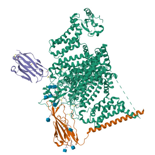
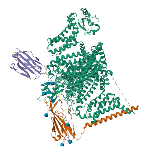
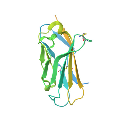
7XM9

Cryo-EM structure of human NaV1.7/beta1/beta2-XEN907


Experimental Data Snapshot

  • Method: ELECTRON MICROSCOPY
  • Resolution: 3.22 Å
  • Aggregation State: PARTICLE 
  • Reconstruction Method: SINGLE PARTICLE 

wwPDB Validation   3D Report Full Report


This is version 1.3 of the entry. See complete history


Literature

Structural basis for Na V 1.7 inhibition by pore blockers.

Zhang, J.Shi, Y.Huang, Z.Li, Y.Yang, B.Gong, J.Jiang, D.

(2022) Nat Struct Mol Biol 29: 1208-1216

  • DOI: https://doi.org/10.1038/s41594-022-00860-1
  • Primary Citation of Related Structures:  
    7XM9, 7XMF, 7XMG

  • PubMed Abstract: 

    Voltage-gated sodium channel Na V 1.7 plays essential roles in pain and odor perception. Na V 1.7 variants cause pain disorders. Accordingly, Na V 1.7 has elicited extensive attention in developing new analgesics. Here we present cryo-EM structures of human Na V 1.7/β1/β2 complexed with inhibitors XEN907, TC-N1752 and Na V 1.7-IN2, explaining specific binding sites and modulation mechanism for the pore blockers. These inhibitors bind in the central cavity blocking ion permeation, but engage different parts of the cavity wall. XEN907 directly causes α- to π-helix transition of DIV-S6 helix, which tightens the fast inactivation gate. TC-N1752 induces π-helix transition of DII-S6 helix mediated by a conserved asparagine on DIII-S6, which closes the activation gate. Na V 1.7-IN2 serves as a pore blocker without causing conformational change. Electrophysiological results demonstrate that XEN907 and TC-N1752 stabilize Na V 1.7 in inactivated state and delay the recovery from inactivation. Our results provide structural framework for Na V 1.7 modulation by pore blockers, and important implications for developing subtype-selective analgesics.


  • Organizational Affiliation

    Laboratory of Soft Matter Physics, Institute of Physics, Chinese Academy of Sciences, Beijing, China.


Macromolecules
Find similar proteins by:  (by identity cutoff)  |  3D Structure
Entity ID: 1
MoleculeChains Sequence LengthOrganismDetailsImage
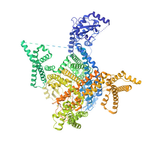
Isoform 3 of Sodium channel protein type 9 subunit alpha,Green fluorescent protein2,250Homo sapiensAequorea victoria
This entity is chimeric
Mutation(s): 1 
Gene Names: SCN9ANENAgfp
Membrane Entity: Yes 
UniProt & NIH Common Fund Data Resources
Find proteins for Q15858 (Homo sapiens)
Explore Q15858 
Go to UniProtKB:  Q15858
PHAROS:  Q15858
GTEx:  ENSG00000169432 
Entity Groups  
Sequence Clusters30% Identity50% Identity70% Identity90% Identity95% Identity100% Identity
UniProt GroupQ15858
Glycosylation
Glycosylation Sites: 3Go to GlyGen: Q15858-1
Sequence Annotations
Expand
  • Reference Sequence
Find similar proteins by:  (by identity cutoff)  |  3D Structure
Entity ID: 2
MoleculeChains Sequence LengthOrganismDetailsImage
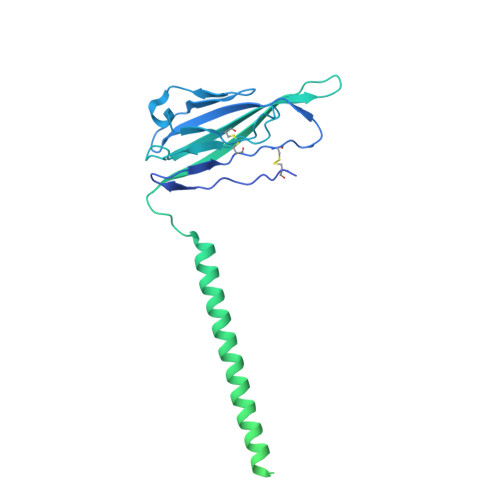
Sodium channel subunit beta-1,Green fluorescent protein481Homo sapiensAequorea victoria
This entity is chimeric
Mutation(s): 0 
Gene Names: SCN1Bgfp
Membrane Entity: Yes 
UniProt & NIH Common Fund Data Resources
Find proteins for Q07699 (Homo sapiens)
Explore Q07699 
Go to UniProtKB:  Q07699
PHAROS:  Q07699
GTEx:  ENSG00000105711 
Entity Groups  
Sequence Clusters30% Identity50% Identity70% Identity90% Identity95% Identity100% Identity
UniProt GroupQ07699
Glycosylation
Glycosylation Sites: 4Go to GlyGen: Q07699-1
Sequence Annotations
Expand
  • Reference Sequence
Find similar proteins by:  (by identity cutoff)  |  3D Structure
Entity ID: 3
MoleculeChains Sequence LengthOrganismDetailsImage
Sodium channel subunit beta-2215Homo sapiensMutation(s): 0 
Gene Names: SCN2B
Membrane Entity: Yes 
UniProt & NIH Common Fund Data Resources
Find proteins for O60939 (Homo sapiens)
Explore O60939 
Go to UniProtKB:  O60939
PHAROS:  O60939
GTEx:  ENSG00000149575 
Entity Groups  
Sequence Clusters30% Identity50% Identity70% Identity90% Identity95% Identity100% Identity
UniProt GroupO60939
Sequence Annotations
Expand
  • Reference Sequence
Oligosaccharides

Help

Entity ID: 4
MoleculeChains Length2D Diagram Glycosylation3D Interactions
2-acetamido-2-deoxy-beta-D-glucopyranose-(1-4)-2-acetamido-2-deoxy-beta-D-glucopyranose
D, E
2N-Glycosylation
Glycosylation Resources
GlyTouCan:  G42666HT
GlyCosmos:  G42666HT
GlyGen:  G42666HT
Small Molecules
Ligands 3 Unique
IDChains Name / Formula / InChI Key2D Diagram3D Interactions
6OU
Query on 6OU

Download Ideal Coordinates CCD File 
H [auth A][(2~{R})-1-[2-azanylethoxy(oxidanyl)phosphoryl]oxy-3-hexadecanoyloxy-propan-2-yl] (~{Z})-octadec-9-enoate
C39 H76 N O8 P
FHQVHHIBKUMWTI-OTMQOFQLSA-N
G2E (Subject of Investigation/LOI)
Query on G2E

Download Ideal Coordinates CCD File 
G [auth A](7~{R})-1'-pentylspiro[6~{H}-furo[3,2-f][1,3]benzodioxole-7,3'-indole]-2'-one
C21 H21 N O4
PHMRUZIIERITEP-OAQYLSRUSA-N
NAG
Query on NAG

Download Ideal Coordinates CCD File 
F [auth A],
I [auth B],
J [auth B],
K [auth B],
L [auth B]
2-acetamido-2-deoxy-beta-D-glucopyranose
C8 H15 N O6
OVRNDRQMDRJTHS-FMDGEEDCSA-N
Binding Affinity Annotations 
IDSourceBinding Affinity
G2E BindingDB:  7XM9 IC50: 3 (nM) from 1 assay(s)
Experimental Data & Validation

Experimental Data

  • Method: ELECTRON MICROSCOPY
  • Resolution: 3.22 Å
  • Aggregation State: PARTICLE 
  • Reconstruction Method: SINGLE PARTICLE 

Structure Validation

View Full Validation Report


Entry History & Funding Information

Deposition Data


Funding OrganizationLocationGrant Number
National Natural Science Foundation of China (NSFC)China31971134

Revision History  (Full details and data files)

  • Version 1.0: 2022-11-30
    Type: Initial release
  • Version 1.1: 2022-12-07
    Changes: Database references
  • Version 1.2: 2022-12-28
    Changes: Database references
  • Version 1.3: 2024-10-30
    Changes: Data collection, Structure summary