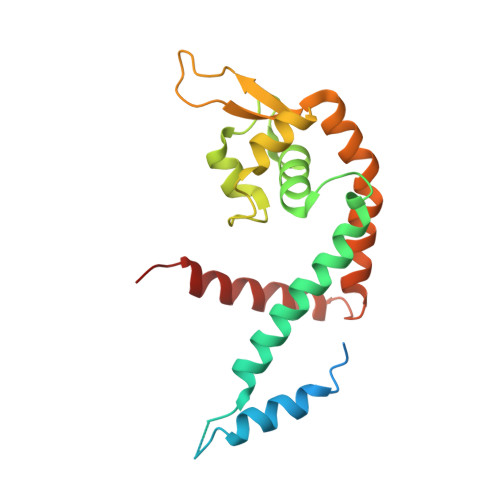
The structure of Deinococcus radiodurans transcriptional regulator HucR retold with the urate bound.
Rho, S., Jung, W., Park, J.K., Choi, M.H., Kim, M., Kim, J., Byun, J., Park, T., Lee, B.I., Wilkinson, S.P., Park, S.(2022) Biochem Biophys Res Commun 615: 63-69
- PubMed: 35605407 Search on PubMed
- DOI: https://doi.org/10.1016/j.bbrc.2022.05.034
- Primary Citation Related Structures:
7XL9 - PubMed Abstract:
HucR is a MarR family protein of Deinococcus radiodurans, which binds tightly to the intergenic region of HucR and the uricase gene to inhibit their expression. Urate (or uric acid) antagonizes the repressor function of HucR by binding to HucR to impede its association with the cognate DNA. The previously reported crystal structure of HucR was without the bound urate showing significant structural homology to other MarR structures. In this paper, we report the crystal structure of HucR determined with the urate bound. However, despite the fact that the urate is found at a site well-known to harbor ligands in other MarR family proteins, the overall HucR structure indicates that no significant change in structure takes place with the urate bound. Structure analysis further suggests that the urate interaction in HucR is mediated by histidine/glutamate side chains and ordered water molecules stabilized by various residues. Such interaction is quite unique compared to other known structural interactions between urate and its binding proteins. Furthermore, structural comparison of the apo- and the urate bound forms allows us to hypothesize that the Trp20-mediated water network in the apo-form stabilizes the proper HucR fold for cognate DNA binding, and that urate binding, also via Trp20, and the consequent reorganization of water molecules in the binding pocket, likely disrupts the DNA binding configuration to result in the attenuated DNA binding.
- School of Systems Biomedical Science and Integrative Institute of Basic Sciences, Soongsil University, Seoul, Republic of Korea.
Organizational Affiliation: