7XF0 | pdb_00007xf0

Crystal strucutre of CasDinG in complex with ATP


Experimental Data Snapshot

  • Method: X-RAY DIFFRACTION
  • Resolution: 3.10 Å
  • R-Value Free: 
    0.261 (Depositor), 0.259 (DCC) 
  • R-Value Work: 
    0.224 (Depositor), 0.224 (DCC) 
  • R-Value Observed: 
    0.225 (Depositor) 

Starting Model: experimental
View more details

wwPDB Validation 3D Report Full Report

Validation slider image for 7XF0

Ligand Structure Quality Assessment 


This is version 1.3 of the entry. See complete history

Literature

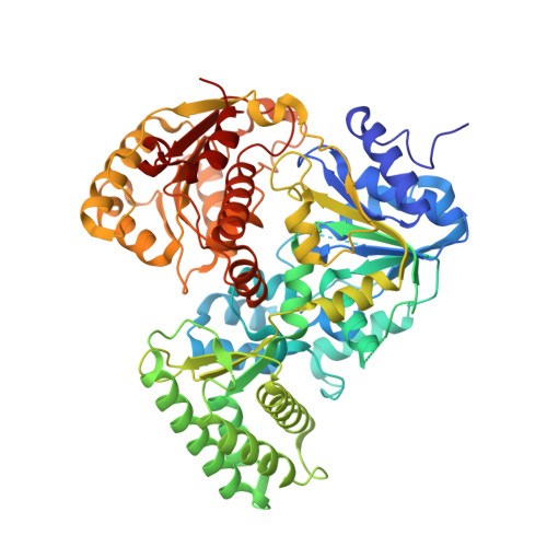
Type IV-A CRISPR-Csf complex: Assembly, dsDNA targeting, and CasDinG recruitment.

Cui, N.Zhang, J.T.Liu, Y.Liu, Y.Liu, X.Y.Wang, C.Huang, H.Jia, N.

(2023) Mol Cell 83: 2493-2508.e5

  • DOI: https://doi.org/10.1016/j.molcel.2023.05.036
  • Primary Citation Related Structures: 
    7XEX, 7XF0, 7XF1, 7XFZ, 7XG0, 7XG2, 7XG3, 7XG4

  • PubMed Abstract: 

    Type IV CRISPR-Cas systems, which are primarily found on plasmids and exhibit a strong plasmid-targeting preference, are the only one of the six known CRISPR-Cas types for which the mechanistic details of their function remain unknown. Here, we provide high-resolution functional snapshots of type IV-A Csf complexes before and after target dsDNA binding, either in the absence or presence of CasDinG, revealing the mechanisms underlying Csf crRNA complex assembly, "DWN" PAM-dependent dsDNA targeting, R-loop formation, and CasDinG recruitment. Furthermore, we establish that CasDinG, a signature DinG family helicase, harbors ssDNA-stimulated ATPase activity and ATP-dependent 5'-3' DNA helicase activity. In addition, we show that CasDinG unwinds the non-target strand (NTS) and target strand (TS) of target dsDNA from the Csf crRNA complex. These molecular details advance our mechanistic understanding of type IV-A CRISPR-Csf function and should enable Csf complexes to be harnessed as genome-engineering tools for biotechnological applications.


  • Organizational Affiliation
    • Department of Biochemistry, School of Medicine, Southern University of Science and Technology, Shenzhen 518055, China.

Macromolecule Content 

  • Total Structure Weight: 205.34 kDa 
  • Atom Count: 13,697 
  • Modeled Residue Count: 1,782 
  • Deposited Residue Count: 1,878 
  • Unique protein chains: 1

Macromolecules

Find similar proteins by:|  3D Structure
Entity ID: 1
MoleculeChains  Sequence LengthOrganismDetailsImage
CasDinGA [auth C],
B [auth A],
C [auth B]
626Pseudomonas aeruginosaMutation(s): 0 
UniProt
Find proteins for A0AA82WNX9 (Pseudomonas aeruginosa)
Explore A0AA82WNX9 
Go to UniProtKB:  A0AA82WNX9
Entity Groups
Sequence Clusters30% Identity50% Identity70% Identity90% Identity95% Identity100% Identity
UniProt GroupA0AA82WNX9
Sequence Annotations
Expand
Reference Sequence

Experimental Data & Validation

Experimental Data

  • Method: X-RAY DIFFRACTION
  • Resolution: 3.10 Å
  • R-Value Free:  0.261 (Depositor), 0.259 (DCC) 
  • R-Value Work:  0.224 (Depositor), 0.224 (DCC) 
  • R-Value Observed: 0.225 (Depositor) 
Space Group: I 1 2 1
Unit Cell:
Length ( Å )Angle ( ˚ )
a = 135.546α = 90
b = 72.095β = 90.84
c = 224.23γ = 90
Software Package:
Software NamePurpose
DIALSdata scaling
PHENIXrefinement

Structure Validation

View Full Validation Report


Ligand Structure Quality Assessment 


Entry History 

& Funding Information

Deposition Data


Funding OrganizationLocationGrant Number
Not funded--

Revision History  (Full details and data files)

  • Version 1.0: 2023-07-26
    Type: Initial release
  • Version 1.1: 2023-08-02
    Changes: Database references
  • Version 1.2: 2024-05-29
    Changes: Data collection
  • Version 1.3: 2026-03-04
    Changes: Refinement description, Structure summary