


Inhibition mechanism of SufS by L-propargylglycine
Nakamura, R., Takahashi, Y., Fujishiro, T.To be published.
Experimental Data Snapshot
Starting Model: experimental
View more details
Entity ID: 1 | |||||
|---|---|---|---|---|---|
| Molecule | Chains | Sequence Length | Organism | Details | Image |
| Cysteine desulfurase SufS | 419 | Bacillus subtilis subsp. subtilis str. 168 | Mutation(s): 0 Gene Names: sufS, csd, yurW, BSU32690 EC: 2.8.1.7 | 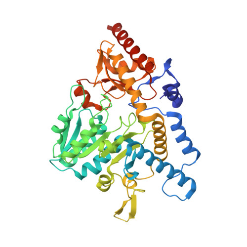 | |
UniProt | |||||
Entity Groups | |||||
| Sequence Clusters | 30% Identity50% Identity70% Identity90% Identity95% Identity100% Identity | ||||
| UniProt Group | O32164 | ||||
Sequence AnnotationsExpand | |||||
Reference Sequence | |||||
| Ligands 3 Unique | |||||
|---|---|---|---|---|---|
| ID | Chains | Name / Formula / InChI Key | 2D Diagram | 3D Interactions | |
| 9YC (Subject of Investigation/LOI) Download:Ideal Coordinates CCD File | B [auth A] | (2~{E})-3-cyano-2-[[2-methyl-3-oxidanyl-5-(phosphonooxymethyl)pyridin-4-yl]methylimino]propanoic acid C12 H14 N3 O7 P RXBCHSOWMGDFEL-XNTDXEJSSA-N |  | ||
| PGE Download:Ideal Coordinates CCD File | E [auth A] | TRIETHYLENE GLYCOL C6 H14 O4 ZIBGPFATKBEMQZ-UHFFFAOYSA-N |  | ||
| PEG Download:Ideal Coordinates CCD File | C [auth A], D [auth A], F [auth A], G [auth A] | DI(HYDROXYETHYL)ETHER C4 H10 O3 MTHSVFCYNBDYFN-UHFFFAOYSA-N |  | ||
| Length ( Å ) | Angle ( ˚ ) |
|---|---|
| a = 92.72 | α = 90 |
| b = 92.72 | β = 90 |
| c = 128.51 | γ = 120 |
| Software Name | Purpose |
|---|---|
| XSCALE | data scaling |
| REFMAC | refinement |
| PDB_EXTRACT | data extraction |
| XDS | data reduction |
| MOLREP | phasing |
| Funding Organization | Location | Grant Number |
|---|---|---|
| Japan Society for the Promotion of Science (JSPS) | Japan | 17K14510 |


