7XEJ | pdb_00007xej

SufS with D-cysteine for 5 min


Experimental Data Snapshot

  • Method: X-RAY DIFFRACTION
  • Resolution: 1.74 Å
  • R-Value Free: 
    0.176 (Depositor), 0.176 (DCC) 
  • R-Value Work: 
    0.145 (Depositor), 0.145 (DCC) 
  • R-Value Observed: 
    0.146 (Depositor) 

Starting Model: experimental
View more details

wwPDB Validation 3D Report Full Report

Validation slider image for 7XEJ

Ligand Structure Quality Assessment 


This is version 1.2 of the entry. See complete history

Literature

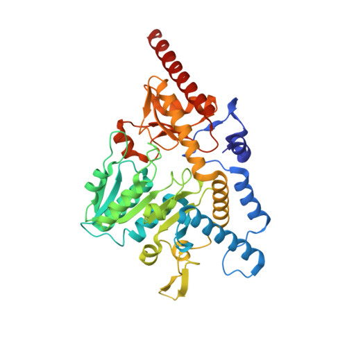
Visualizing thiazolidine ring formation in the reaction of D-cysteine and pyridoxal-5'-phosphate within L-cysteine desulfurase SufS.

Nakamura, R.Fujishiro, T.

(2025) Biochem Biophys Res Commun 754: 151497-151497

  • DOI: https://doi.org/10.1016/j.bbrc.2025.151497
  • Primary Citation Related Structures: 
    7XEJ, 7XEK, 7XEL, 7XEN, 7YB3

  • PubMed Abstract: 

    The reactivity of pyridoxal-5'-phosphate (PLP) with cysteine and its derivatives has been of increasing interest because the corresponding product, a thiazolidine PLP-cysteine adduct, can be formed via PLP-dependent enzymatic and non-enzymatic reactions. Here, we report biochemical and X-ray crystallographic snapshots of thiazolidine formation in reaction of D-cysteine with PLP in SufS, a PLP-dependent L-cysteine desulfurase. By comparing L- and D-penicillamine-bound SufS showing no thiazolidine formation in the crystals with D-cysteine SufS, we proposed a thiazolidine formation mechanism with important factors: the polar environments provided by the carbonyl groups of Ala28-Ala29 and Lys224-mediated base catalysis for the nucleophilic thiolate of D-cysteine.


  • Organizational Affiliation
    • Department of Biochemistry and Molecular Biology, Graduate School of Science and Engineering, Saitama University, Japan.

Macromolecule Content 

  • Total Structure Weight: 47.89 kDa 
  • Atom Count: 3,706 
  • Modeled Residue Count: 413 
  • Deposited Residue Count: 419 
  • Unique protein chains: 1

Macromolecules

Find similar proteins by:|  3D Structure
Entity ID: 1
MoleculeChains  Sequence LengthOrganismDetailsImage
Cysteine desulfurase SufS419Bacillus subtilis subsp. subtilis str. 168Mutation(s): 0 
Gene Names: sufScsdyurWBSU32690
EC: 2.8.1.7
UniProt
Find proteins for O32164 (Bacillus subtilis (strain 168))
Explore O32164 
Go to UniProtKB:  O32164
Entity Groups
Sequence Clusters30% Identity50% Identity70% Identity90% Identity95% Identity100% Identity
UniProt GroupO32164
Sequence Annotations
Expand
Reference Sequence

Small Molecules

Ligands 4 Unique
IDChains Name / Formula / InChI Key2D Diagram3D Interactions
9PV
(Subject of Investigation/LOI)

Query on 9PV


Download:Ideal Coordinates CCD File
B [auth A](2~{S})-2-[(~{E})-[2-methyl-3-oxidanyl-5-(phosphonooxymethyl)pyridin-4-yl]methylideneamino]-3-sulfanyl-propanoic acid
C11 H15 N2 O7 P S
NUTWYOJHALJXFM-RZTFRGLUSA-N
PGE

Query on PGE


Download:Ideal Coordinates CCD File
K [auth A]TRIETHYLENE GLYCOL
C6 H14 O4
ZIBGPFATKBEMQZ-UHFFFAOYSA-N
PEG

Query on PEG


Download:Ideal Coordinates CCD File
D [auth A]
E [auth A]
F [auth A]
G [auth A]
H [auth A]
D [auth A],
E [auth A],
F [auth A],
G [auth A],
H [auth A],
I [auth A],
J [auth A]
DI(HYDROXYETHYL)ETHER
C4 H10 O3
MTHSVFCYNBDYFN-UHFFFAOYSA-N
EDO

Query on EDO


Download:Ideal Coordinates CCD File
C [auth A]1,2-ETHANEDIOL
C2 H6 O2
LYCAIKOWRPUZTN-UHFFFAOYSA-N

Experimental Data & Validation

Experimental Data

  • Method: X-RAY DIFFRACTION
  • Resolution: 1.74 Å
  • R-Value Free:  0.176 (Depositor), 0.176 (DCC) 
  • R-Value Work:  0.145 (Depositor), 0.145 (DCC) 
  • R-Value Observed: 0.146 (Depositor) 
Space Group: P 31 2 1
Unit Cell:
Length ( Å )Angle ( ˚ )
a = 92.9α = 90
b = 92.9β = 90
c = 129.4γ = 120
Software Package:
Software NamePurpose
XSCALEdata scaling
REFMACrefinement
PDB_EXTRACTdata extraction
XDSdata reduction
MOLREPphasing

Structure Validation

View Full Validation Report


Ligand Structure Quality Assessment 


Entry History 

& Funding Information

Deposition Data


Funding OrganizationLocationGrant Number
Japan Society for the Promotion of Science (JSPS)Japan17K14510

Revision History  (Full details and data files)

  • Version 1.0: 2023-04-05
    Type: Initial release
  • Version 1.1: 2023-11-29
    Changes: Data collection, Refinement description
  • Version 1.2: 2025-04-02
    Changes: Database references, Structure summary