7XAX | pdb_00007xax

Crystal structure of SARS coronavirus main protease in complex with Baicalei


Experimental Data Snapshot

  • Method: X-RAY DIFFRACTION
  • Resolution: 2.25 Å
  • R-Value Free: 
    0.280 (Depositor), 0.281 (DCC) 
  • R-Value Work: 
    0.229 (Depositor), 0.229 (DCC) 
  • R-Value Observed: 
    0.232 (Depositor) 

Starting Model: experimental
View more details

wwPDB Validation 3D Report Full Report

Validation slider image for 7XAX

Ligand Structure Quality Assessment 


This is version 1.3 of the entry. See complete history

Literature

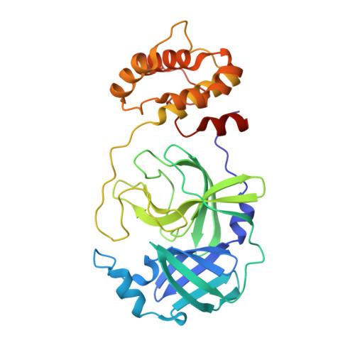
Crystal structure of SARS-CoV 3C-like protease with baicalein.

Feng, J.Li, D.Zhang, J.Yin, X.Li, J.

(2022) Biochem Biophys Res Commun 611: 190-194

  • DOI: https://doi.org/10.1016/j.bbrc.2022.04.086
  • Primary Citation Related Structures: 
    7XAX

  • PubMed Abstract: 

    The 3C-like protease (M pro , 3CL pro ) plays a key role in the replication process in coronaviruses (CoVs). The M pro is an essential enzyme mediates CoVs replication and is a promising target for development of antiviral drugs. Until now, baicalein has been shown the specific activity for SARS-CoV M pro in vitro experiments. In this study, we resolved the SARS-CoV M pro with baicalein by X-ray diffraction at 2.25 Å (PDB code 7XAX), which provided a structural basis for the research and development of baicalein as an anti-CoVs drug.


  • Organizational Affiliation
    • Applied Biology Laboratory, College of Pharmaceutical and Biological Engineering, Shenyang University of Chemical Technology, Shenyang, 110142, China.

Macromolecule Content 

  • Total Structure Weight: 68.02 kDa 
  • Atom Count: 4,435 
  • Modeled Residue Count: 594 
  • Deposited Residue Count: 612 
  • Unique protein chains: 1

Macromolecules

Find similar proteins by:|  3D Structure
Entity ID: 1
MoleculeChains  Sequence LengthOrganismDetailsImage
3C-like proteinase nsp5
A, B
306Severe acute respiratory syndrome-related coronavirusMutation(s): 0 
EC: 3.4.22.69
UniProt
Find proteins for P0C6X7 (Severe acute respiratory syndrome coronavirus)
Explore P0C6X7 
Go to UniProtKB:  P0C6X7
Entity Groups
Sequence Clusters30% Identity50% Identity70% Identity90% Identity95% Identity100% Identity
UniProt GroupP0C6X7
Sequence Annotations
Expand
Reference Sequence

Small Molecules

Ligands 1 Unique
IDChains Name / Formula / InChI Key2D Diagram3D Interactions
3WL
(Subject of Investigation/LOI)

Query on 3WL


Download:Ideal Coordinates CCD File
C [auth A]5,6,7-trihydroxy-2-phenyl-4H-chromen-4-one
C15 H10 O5
FXNFHKRTJBSTCS-UHFFFAOYSA-N

Experimental Data & Validation

Experimental Data

  • Method: X-RAY DIFFRACTION
  • Resolution: 2.25 Å
  • R-Value Free:  0.280 (Depositor), 0.281 (DCC) 
  • R-Value Work:  0.229 (Depositor), 0.229 (DCC) 
  • R-Value Observed: 0.232 (Depositor) 
Space Group: P 1
Unit Cell:
Length ( Å )Angle ( ˚ )
a = 55.51α = 90.951
b = 60.55β = 102.715
c = 68.28γ = 108.649
Software Package:
Software NamePurpose
PHENIXrefinement
XDSdata scaling
PHENIXphasing

Structure Validation

View Full Validation Report


Ligand Structure Quality Assessment 


Entry History 

& Funding Information

Deposition Data


Funding OrganizationLocationGrant Number
Not funded--

Revision History  (Full details and data files)

  • Version 1.0: 2023-03-22
    Type: Initial release
  • Version 1.1: 2023-11-29
    Changes: Data collection, Refinement description
  • Version 1.2: 2024-10-02
    Changes: Database references
  • Version 1.3: 2025-09-17
    Changes: Advisory, Derived calculations, Structure summary