7X6Y

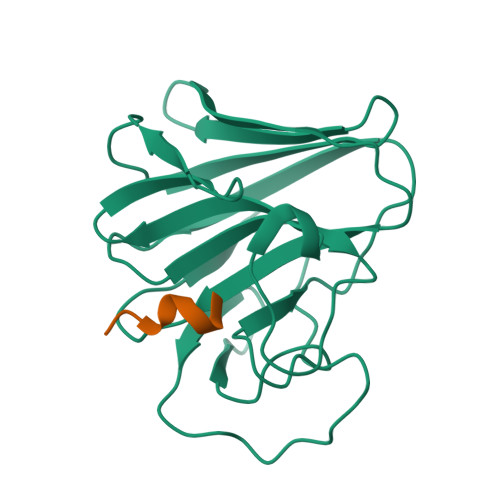
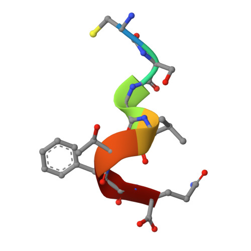
TRIM7 in complex with C-terminal peptide of NSP5


Experimental Data Snapshot

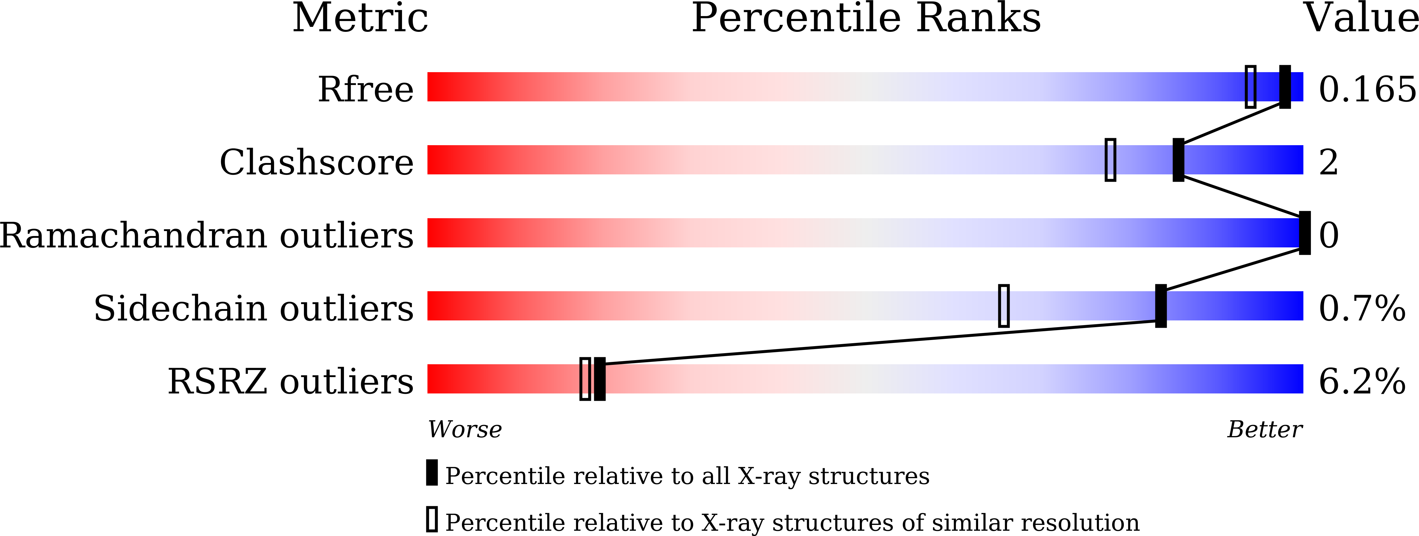
  • Method: X-RAY DIFFRACTION
  • Resolution: 1.39 Å
  • R-Value Free: 0.165 
  • R-Value Work: 0.143 
  • R-Value Observed: 0.144 

Starting Model: experimental
View more details

wwPDB Validation   3D Report Full Report


This is version 1.3 of the entry. See complete history


Literature

A C-terminal glutamine recognition mechanism revealed by E3 ligase TRIM7 structures.

Liang, X.Xiao, J.Li, X.Liu, Y.Lu, Y.Wen, Y.Li, Z.Che, X.Ma, Y.Zhang, X.Zhang, Y.Jian, D.Wang, P.Xuan, C.Yu, G.Li, L.Zhang, H.

(2022) Nat Chem Biol 18: 1214-1223

  • DOI: https://doi.org/10.1038/s41589-022-01128-x
  • Primary Citation of Related Structures:  
    7W0Q, 7W0S, 7W0T, 7X6Y, 7X6Z, 7X70

  • PubMed Abstract: 

    The E3 ligase TRIM7 has emerged as a critical player in viral infection and pathogenesis. However, the mechanism governing the TRIM7-substrate association remains to be defined. Here we report the crystal structures of TRIM7 in complex with 2C peptides of human enterovirus. Structure-guided studies reveal the C-terminal glutamine residue of 2C as the primary determinant for TRIM7 binding. Leveraged by this finding, we identify norovirus and SARS-CoV-2 proteins, and physiological proteins, as new TRIM7 substrates. Crystal structures of TRIM7 in complex with multiple peptides derived from SARS-CoV-2 proteins display the same glutamine-end recognition mode. Furthermore, TRIM7 could trigger the ubiquitination and degradation of these substrates, possibly representing a new Gln/C-degron pathway. Together, these findings unveil a common recognition mode by TRIM7, providing the foundation for further mechanistic characterization of antiviral and cellular functions of TRIM7.


  • Organizational Affiliation

    Key Laboratory of Immune Microenvironment and Disease (Ministry of Education), The Province and Ministry Co-sponsored Collaborative Innovation Center for Medical Epigenetics, Haihe Laboratory of Cell Ecosystem, Tianjin Institute of Immunology, Department of Biochemistry and Molecular Biology, School of Basic Medical Sciences, Tianjin Medical University, Tianjin, China.


Macromolecules
Find similar proteins by:  (by identity cutoff)  |  3D Structure
Entity ID: 1
MoleculeChains Sequence LengthOrganismDetailsImage
E3 ubiquitin-protein ligase TRIM7174Homo sapiensMutation(s): 0 
Gene Names: TRIM7GNIPRNF90
EC: 2.3.2.27
UniProt & NIH Common Fund Data Resources
Find proteins for Q9C029 (Homo sapiens)
Explore Q9C029 
Go to UniProtKB:  Q9C029
PHAROS:  Q9C029
GTEx:  ENSG00000146054 
Entity Groups  
Sequence Clusters30% Identity50% Identity70% Identity90% Identity95% Identity100% Identity
UniProt GroupQ9C029
Sequence Annotations
Expand
  • Reference Sequence

Find similar proteins by:  Sequence   |   3D Structure  

Entity ID: 2
MoleculeChains Sequence LengthOrganismDetailsImage
peptide7Severe acute respiratory syndrome coronavirus 2Mutation(s): 0 
UniProt
Find proteins for P0DTD1 (Severe acute respiratory syndrome coronavirus 2)
Explore P0DTD1 
Go to UniProtKB:  P0DTD1
Entity Groups  
UniProt GroupP0DTD1
Sequence Annotations
Expand
  • Reference Sequence
Experimental Data & Validation

Experimental Data

  • Method: X-RAY DIFFRACTION
  • Resolution: 1.39 Å
  • R-Value Free: 0.165 
  • R-Value Work: 0.143 
  • R-Value Observed: 0.144 
  • Space Group: P 65
Unit Cell:
Length ( Å )Angle ( ˚ )
a = 79.915α = 90
b = 79.915β = 90
c = 53.083γ = 120
Software Package:
Software NamePurpose
PHENIXrefinement
autoPROCdata reduction
autoPROCdata scaling
PHENIXphasing

Structure Validation

View Full Validation Report


Entry History & Funding Information

Deposition Data


Funding OrganizationLocationGrant Number
Other government--

Revision History  (Full details and data files)

  • Version 1.0: 2022-08-10
    Type: Initial release
  • Version 1.1: 2022-10-26
    Changes: Database references
  • Version 1.2: 2022-11-09
    Changes: Database references
  • Version 1.3: 2023-11-29
    Changes: Data collection, Refinement description