7X1M | pdb_00007x1m

The complex structure of Omicron BA.1 RBD with BD604, S309,and S304


Experimental Data Snapshot

  • Method: ELECTRON MICROSCOPY
  • Resolution: 2.74 Å
  • Aggregation State: PARTICLE 
  • Reconstruction Method: SINGLE PARTICLE 

wwPDB Validation 3D Report Full Report

Validation slider image for 7X1M

This is version 1.3 of the entry. See complete history

Literature

Atlas of currently available human neutralizing antibodies against SARS-CoV-2 and escape by Omicron sub-variants BA.1/BA.1.1/BA.2/BA.3.

Huang, M.Wu, L.Zheng, A.Xie, Y.He, Q.Rong, X.Han, P.Du, P.Han, P.Zhang, Z.Zhao, R.Jia, Y.Li, L.Bai, B.Hu, Z.Hu, S.Niu, S.Hu, Y.Liu, H.Liu, B.Cui, K.Li, W.Zhao, X.Liu, K.Qi, J.Wang, Q.Gao, G.F.

(2022) Immunity 55: 1501-1514.e3

  • DOI: https://doi.org/10.1016/j.immuni.2022.06.005
  • Primary Citation Related Structures: 
    7X1M

  • PubMed Abstract: 

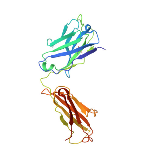
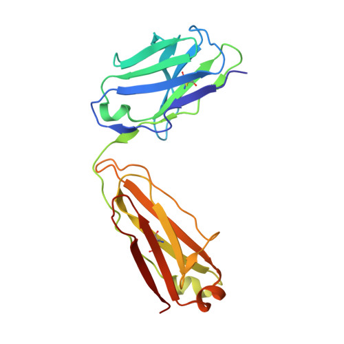
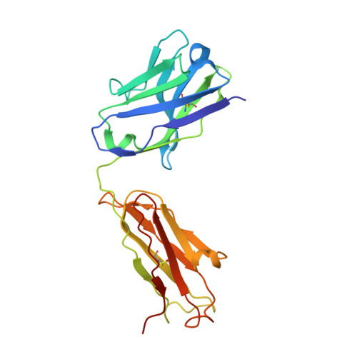
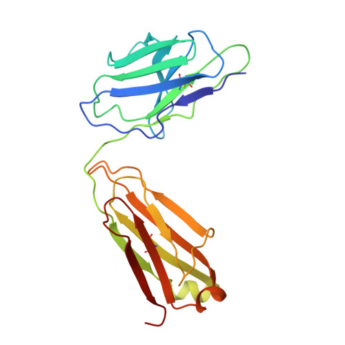
    SARS-CoV-2 Omicron variant has presented significant challenges to current antibodies and vaccines. Herein, we systematically compared the efficacy of 50 human monoclonal antibodies (mAbs), covering the seven identified epitope classes of the SARS-CoV-2 RBD, against Omicron sub-variants BA.1, BA.1.1, BA.2, and BA.3. Binding and pseudovirus-based neutralizing assays revealed that 37 of the 50 mAbs lost neutralizing activities, whereas the others displayed variably decreased activities against the four Omicron sub-variants. BA.2 was found to be more sensitive to RBD-5 antibodies than the other sub-variants. Furthermore, a quaternary complex structure of BA.1 RBD with three mAbs showing different neutralizing potencies against Omicron provided a basis for understanding the immune evasion of Omicron sub-variants and revealed the lack of G446S mutation accounting for the sensitivity of BA.2 to RBD-5 mAbs. Our results may guide the application of the available mAbs and facilitate the development of universal therapeutic antibodies and vaccines against COVID-19.


  • Organizational Affiliation
    • School of Life Science, Division of Life Sciences and Medicine, University of Science and Technology of China, Hefei, Anhui 230027, China; CAS Key Laboratory of Pathogen Microbiology and Immunology, Institute of Microbiology, Chinese Academy of Sciences, Beijing 100101, China.

Macromolecule Content 

  • Total Structure Weight: 165.88 kDa 
  • Atom Count: 9,908 
  • Modeled Residue Count: 1,291 
  • Deposited Residue Count: 1,527 
  • Unique protein chains: 7

Macromolecules

Find similar proteins by:|  3D Structure
Entity ID: 1
MoleculeChains  Sequence LengthOrganismDetailsImage
S309 Fab heavy chain230Homo sapiensMutation(s): 0 
Find similar proteins by:|  3D Structure
Entity ID: 2
MoleculeChains  Sequence LengthOrganismDetailsImage
S309 Fab light chain214Homo sapiensMutation(s): 0 
Find similar proteins by:|  3D Structure
Entity ID: 3
MoleculeChains  Sequence LengthOrganismDetailsImage
BD-604 Fab heavy chain222Homo sapiensMutation(s): 0 
Find similar proteins by:|  3D Structure
Entity ID: 4
MoleculeChains  Sequence LengthOrganismDetailsImage
BD-604 Fab light chain214Homo sapiensMutation(s): 0 
Find similar proteins by:|  3D Structure
Entity ID: 5
MoleculeChains  Sequence LengthOrganismDetailsImage
S304 Fab haavy chainE [auth H]223Homo sapiensMutation(s): 0 
Find similar proteins by:|  3D Structure
Entity ID: 6
MoleculeChains  Sequence LengthOrganismDetailsImage
S304 Fab light chainF [auth L]215Homo sapiensMutation(s): 0 
Find similar proteins by:|  3D Structure
Entity ID: 7
MoleculeChains  Sequence LengthOrganismDetailsImage
Spike protein S1G [auth M]209Severe acute respiratory syndrome coronavirus 2Mutation(s): 15 
Gene Names: S2
UniProt
Find proteins for P0DTC2 (Severe acute respiratory syndrome coronavirus 2)
Explore P0DTC2 
Go to UniProtKB:  P0DTC2
Entity Groups
Sequence Clusters30% Identity50% Identity70% Identity90% Identity95% Identity100% Identity
UniProt GroupP0DTC2
Glycosylation
Glycosylation Sites: 1Go to GlyGen: P0DTC2-1
Sequence Annotations
Expand
Reference Sequence

Oligosaccharides

Help  
Entity ID: 8
MoleculeChains Length2D Diagram GlycosylationD Interactions
2-acetamido-2-deoxy-beta-D-glucopyranose-(1-4)-[alpha-L-fucopyranose-(1-6)]2-acetamido-2-deoxy-beta-D-glucopyranoseH [auth E]3N-Glycosylation
Glycosylation Resources
GlyTouCan: G21290RB
GlyCosmos: G21290RB
GlyGen: G21290RB

Experimental Data & Validation

Experimental Data

  • Method: ELECTRON MICROSCOPY
  • Resolution: 2.74 Å
  • Aggregation State: PARTICLE 
  • Reconstruction Method: SINGLE PARTICLE 
EM Software:
TaskSoftware PackageVersion
MODEL REFINEMENTPHENIX

Structure Validation

View Full Validation Report


Entry History 

& Funding Information

Deposition Data


Funding OrganizationLocationGrant Number
Chinese Academy of SciencesChinaYSBR-010

Revision History  (Full details and data files)

  • Version 1.0: 2022-07-06
    Type: Initial release
  • Version 1.1: 2023-07-19
    Changes: Database references
  • Version 1.2: 2024-10-30
    Changes: Data collection, Structure summary
  • Version 1.3: 2025-07-02
    Changes: Data collection