7X08 | pdb_00007x08

S protein of SARS-CoV-2 in complex with 2G1


Experimental Data Snapshot

  • Method: ELECTRON MICROSCOPY
  • Resolution: 2.70 Å
  • Aggregation State: PARTICLE 
  • Reconstruction Method: SINGLE PARTICLE 

wwPDB Validation 3D Report Full Report

Validation slider image for 7X08

This is version 2.1 of the entry. See complete history

Literature

Broad ultra-potent neutralization of SARS-CoV-2 variants by monoclonal antibodies specific to the tip of RBD.

Ma, H.Guo, Y.Tang, H.Tseng, C.K.Wang, L.Zong, H.Wang, Z.He, Y.Chang, Y.Wang, S.Huang, H.Ke, Y.Yuan, Y.Wu, M.Zhang, Y.Drelich, A.Kempaiah, K.R.Peng, B.H.Wang, A.Yang, K.Yin, H.Liu, J.Yue, Y.Xu, W.Zhu, S.Ji, T.Zhang, X.Wang, Z.Li, G.Liu, G.Song, J.Mu, L.Xiang, Z.Song, Z.Chen, H.Bian, Y.Zhang, B.Chen, H.Zhang, J.Liao, Y.Zhang, L.Yang, L.Chen, Y.Gilly, J.Xiao, X.Han, L.Jiang, H.Xie, Y.Zhou, Q.Zhu, J.

(2022) Cell Discov 8: 16-16

  • DOI: https://doi.org/10.1038/s41421-022-00381-7
  • Primary Citation Related Structures: 
    7X08

  • PubMed Abstract: 

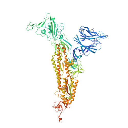
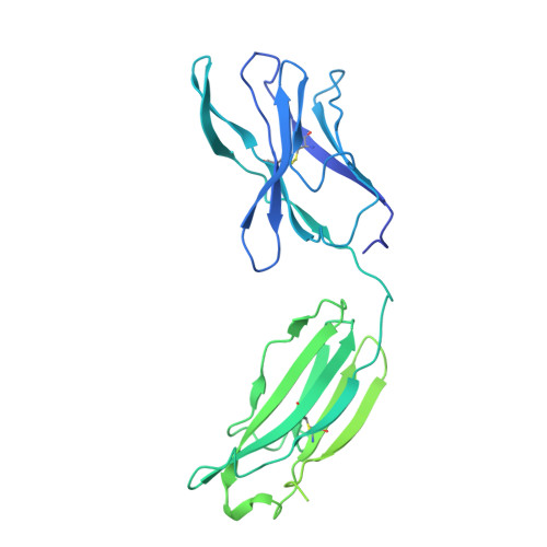
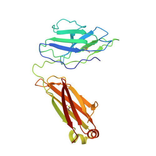
    Severe acute respiratory syndrome coronavirus 2 (SARS-CoV-2) variants of concern (VOCs) continue to wreak havoc across the globe. Higher transmissibility and immunologic resistance of VOCs bring unprecedented challenges to epidemic extinguishment. Here we describe a monoclonal antibody, 2G1, that neutralizes all current VOCs and has surprising tolerance to mutations adjacent to or within its interaction epitope. Cryo-electron microscopy structure showed that 2G1 bound to the tip of receptor binding domain (RBD) of spike protein with small contact interface but strong hydrophobic effect, which resulted in nanomolar to sub-nanomolar affinities to spike proteins. The epitope of 2G1 on RBD partially overlaps with angiotensin converting enzyme 2 (ACE2) interface, which enables 2G1 to block interaction between RBD and ACE2. The narrow binding epitope but high affinity bestow outstanding therapeutic efficacy upon 2G1 that neutralized VOCs with sub-nanomolar half maximal inhibitory concentration in vitro. In SARS-CoV-2, Beta or Delta variant-challenged transgenic mice and rhesus macaque models, 2G1 protected animals from clinical illness and eliminated viral burden, without serious impact to animal safety. Mutagenesis experiments suggest that 2G1 is potentially capable of dealing with emerging SARS-CoV-2 variants in the future. This report characterized the therapeutic antibodies specific to the tip of spike against SARS-CoV-2 variants and highlights the potential clinical applications as well as for developing vaccine and cocktail therapy.


  • Organizational Affiliation
    • Engineering Research Center of Cell and Therapeutic Antibody, Ministry of Education, China; School of Pharmacy, Shanghai Jiao Tong University, Shanghai, China.

Macromolecule Content 

  • Total Structure Weight: 661.41 kDa 
  • Atom Count: 36,396 
  • Modeled Residue Count: 4,572 
  • Deposited Residue Count: 5,853 
  • Unique protein chains: 3

Macromolecules

Find similar proteins by:|  3D Structure
Entity ID: 1
MoleculeChains  Sequence LengthOrganismDetailsImage
Spike glycoprotein
A, B, C
1,283Severe acute respiratory syndrome coronavirusMutation(s): 6 
Gene Names: S2
UniProt
Find proteins for P0DTC2 (Severe acute respiratory syndrome coronavirus 2)
Explore P0DTC2 
Go to UniProtKB:  P0DTC2
Entity Groups
Sequence Clusters30% Identity50% Identity70% Identity90% Identity95% Identity100% Identity
UniProt GroupP0DTC2
Glycosylation
Glycosylation Sites: 18Go to GlyGen: P0DTC2-1
Sequence Annotations
Expand
Reference Sequence
Find similar proteins by:|  3D Structure
Entity ID: 2
MoleculeChains  Sequence LengthOrganismDetailsImage
heavy chain of 2G1D [auth H],
F [auth I],
H [auth J]
452Homo sapiensMutation(s): 0 
Find similar proteins by:|  3D Structure
Entity ID: 3
MoleculeChains  Sequence LengthOrganismDetailsImage
light chain of 2G1E [auth L],
G [auth M],
I [auth N]
216Homo sapiensMutation(s): 0 

Small Molecules

Ligands 2 Unique
IDChains Name / Formula / InChI Key2D Diagram3D Interactions
EIC
(Subject of Investigation/LOI)

Query on EIC


Download:Ideal Coordinates CCD File
DB [auth B],
RA [auth A],
SA [auth A]
LINOLEIC ACID
C18 H32 O2
OYHQOLUKZRVURQ-HZJYTTRNSA-N
NAG
(Subject of Investigation/LOI)

Query on NAG


Download:Ideal Coordinates CCD File
AB [auth B]
BB [auth B]
CB [auth B]
EB [auth C]
FB [auth C]
AB [auth B],
BB [auth B],
CB [auth B],
EB [auth C],
FB [auth C],
GB [auth C],
HA [auth A],
HB [auth C],
IA [auth A],
IB [auth C],
JA [auth A],
JB [auth C],
KA [auth A],
KB [auth C],
LA [auth A],
LB [auth C],
MA [auth A],
MB [auth C],
NA [auth A],
NB [auth C],
OA [auth A],
PA [auth A],
QA [auth A],
TA [auth B],
UA [auth B],
VA [auth B],
WA [auth B],
XA [auth B],
YA [auth B],
ZA [auth B]
2-acetamido-2-deoxy-beta-D-glucopyranose
C8 H15 N O6
OVRNDRQMDRJTHS-FMDGEEDCSA-N

Experimental Data & Validation

Experimental Data

  • Method: ELECTRON MICROSCOPY
  • Resolution: 2.70 Å
  • Aggregation State: PARTICLE 
  • Reconstruction Method: SINGLE PARTICLE 
EM Software:
TaskSoftware PackageVersion
RECONSTRUCTIONRELION3.0.6

Structure Validation

View Full Validation Report


Entry History 

& Funding Information

Deposition Data


Funding OrganizationLocationGrant Number
National Natural Science Foundation of China (NSFC)China31971123

Revision History  (Full details and data files)

  • Version 1.0: 2022-03-09
    Type: Initial release
  • Version 2.0: 2023-11-15
    Changes: Atomic model, Data collection, Derived calculations
  • Version 2.1: 2024-11-06
    Changes: Data collection, Structure summary