7WY2 | pdb_00007wy2

Structure of the Oxomolybdenum Mesoporphyrin IX-Reconstituted CYP102A1 F87A Mutant Haem Domain with N-Enanthyl-L-Prolyl-L-Phenylalanine in complex with Styrene


Experimental Data Snapshot

  • Method: X-RAY DIFFRACTION
  • Resolution: 1.45 Å
  • R-Value Free: 
    0.185 (Depositor), 0.204 (DCC) 
  • R-Value Work: 
    0.148 (Depositor) 
  • R-Value Observed: 
    0.150 (Depositor) 

Starting Model: experimental
View more details

wwPDB Validation 3D Report Full Report

Validation slider image for 7WY2

Ligand Structure Quality Assessment 


This is version 1.4 of the entry. See complete history

Literature

A Compound I Mimic Reveals the Transient Active Species of a Cytochrome P450 Enzyme: Insight into the Stereoselectivity of P450-Catalysed Oxidations.

Suzuki, K.Stanfield, J.K.Omura, K.Shisaka, Y.Ariyasu, S.Kasai, C.Aiba, Y.Sugimoto, H.Shoji, O.

(2023) Angew Chem Int Ed Engl 62: e202215706-e202215706

  • DOI: https://doi.org/10.1002/anie.202215706
  • Primary Citation Related Structures: 
    7WY1, 7WY2, 7WY3, 7WY4

  • PubMed Abstract: 

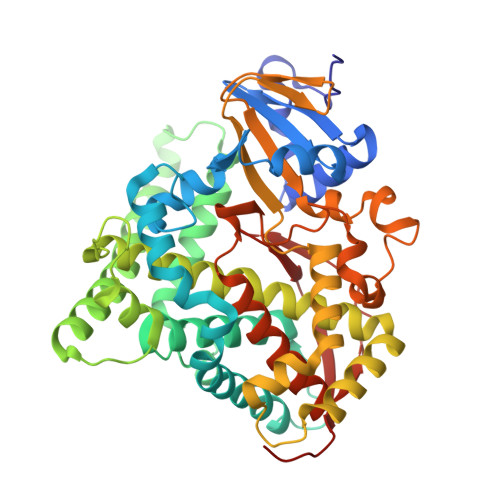
    Catching the structure of cytochrome P450 enzymes in flagrante is crucial for the development of P450 biocatalysts, as most structures collected are found trapped in a precatalytic conformation. At the heart of P450 catalysis lies Cpd I, a short-lived, highly reactive intermediate, whose recalcitrant nature has thwarted most attempts at capturing catalytically relevant poses of P450s. We report the crystal structure of P450BM3 mimicking the state in the precise moment preceding epoxidation, which is in perfect agreement with the experimentally observed stereoselectivity. This structure was attained by incorporation of the stable Cpd I mimic oxomolybdenum mesoporphyrin IX into P450BM3 in the presence of styrene. The orientation of styrene to the Mo-oxo species in the crystal structures sheds light onto the dynamics involved in the rotation of styrene to present its vinyl group to Cpd I. This method serves as a powerful tool for predicting and modelling the stereoselectivity of P450 reactions.


  • Organizational Affiliation
    • Department of Chemistry, Graduate School of Science, Nagoya University Furo-cho, Chikusa-ku, Nagoya, 464-8602, Japan.

Macromolecule Content 

  • Total Structure Weight: 108.09 kDa 
  • Atom Count: 8,696 
  • Modeled Residue Count: 908 
  • Deposited Residue Count: 912 
  • Unique protein chains: 1

Macromolecules

Find similar proteins by:|  3D Structure
Entity ID: 1
MoleculeChains  Sequence LengthOrganismDetailsImage
Bifunctional cytochrome P450/NADPH--P450 reductase
A, B
456Priestia megateriumMutation(s): 1 
Gene Names: BTA37_15100
EC: 1.14.14.1 (PDB Primary Data), 1.6.2.4 (PDB Primary Data)
UniProt
Find proteins for P14779 (Priestia megaterium (strain ATCC 14581 / DSM 32 / CCUG 1817 / JCM 2506 / NBRC 15308 / NCIMB 9376 / NCTC 10342 / NRRL B-14308 / VKM B-512 / Ford 19))
Explore P14779 
Go to UniProtKB:  P14779
Entity Groups
Sequence Clusters30% Identity50% Identity70% Identity90% Identity95% Identity100% Identity
UniProt GroupP14779
Sequence Annotations
Expand
Reference Sequence

Small Molecules

Ligands 5 Unique
IDChains Name / Formula / InChI Key2D Diagram3D Interactions
MI9
(Subject of Investigation/LOI)

Query on MI9


Download:Ideal Coordinates CCD File
C [auth A],
O [auth B]
Oxomolybdenum Mesoporphyrin IX
C34 H36 Mo N4 O5
BVPDTXBFNWLJAZ-HXFTUNQESA-L
D0L

Query on D0L


Download:Ideal Coordinates CCD File
D [auth A],
P [auth B]
(2S)-2-[[(2S)-1-heptylpyrrolidin-2-yl]carbonylamino]-3-phenyl-propanoic acid
C21 H32 N2 O3
UZXMNGCHFSENBI-OALUTQOASA-N
SYN

Query on SYN


Download:Ideal Coordinates CCD File
E [auth A],
Q [auth B]
ethenylbenzene
C8 H8
PPBRXRYQALVLMV-UHFFFAOYSA-N
GOL

Query on GOL


Download:Ideal Coordinates CCD File
F [auth A]
G [auth A]
H [auth A]
I [auth A]
J [auth A]
F [auth A],
G [auth A],
H [auth A],
I [auth A],
J [auth A],
K [auth A],
L [auth A],
M [auth A],
R [auth B],
S [auth B],
T [auth B],
U [auth B],
V [auth B],
W [auth B]
GLYCEROL
C3 H8 O3
PEDCQBHIVMGVHV-UHFFFAOYSA-N
CL

Query on CL


Download:Ideal Coordinates CCD File
N [auth A],
X [auth B]
CHLORIDE ION
Cl
VEXZGXHMUGYJMC-UHFFFAOYSA-M

Experimental Data & Validation

Experimental Data

  • Method: X-RAY DIFFRACTION
  • Resolution: 1.45 Å
  • R-Value Free:  0.185 (Depositor), 0.204 (DCC) 
  • R-Value Work:  0.148 (Depositor) 
  • R-Value Observed: 0.150 (Depositor) 
Space Group: P 21 21 21
Unit Cell:
Length ( Å )Angle ( ˚ )
a = 59.2α = 90
b = 127.31β = 90
c = 148.77γ = 90
Software Package:
Software NamePurpose
XSCALEdata scaling
REFMACrefinement
PDB_EXTRACTdata extraction
XDSdata reduction
MOLREPphasing

Structure Validation

View Full Validation Report


Ligand Structure Quality Assessment 


Entry History 

& Funding Information

Deposition Data


Funding OrganizationLocationGrant Number
Japan Science and TechnologyJapanJPMJCR15P3
Japan Society for the Promotion of Science (JSPS)JapanJP15H05806
Japan Society for the Promotion of Science (JSPS)JapanJP18J23340
Japan Society for the Promotion of Science (JSPS)JapanJP18J15250

Revision History  (Full details and data files)

  • Version 1.0: 2023-01-04
    Type: Initial release
  • Version 1.1: 2023-02-15
    Changes: Database references
  • Version 1.2: 2023-03-29
    Changes: Database references
  • Version 1.3: 2023-05-03
    Changes: Database references
  • Version 1.4: 2023-11-29
    Changes: Data collection, Refinement description