7WWX | pdb_00007wwx

Crystal structure of Herbaspirillum huttiense L-arabinose 1-dehydrogenase (NAD bound form)


Experimental Data Snapshot

  • Method: X-RAY DIFFRACTION
  • Resolution: 1.36 Å
  • R-Value Free: 
    0.192 (Depositor), 0.192 (DCC) 
  • R-Value Work: 
    0.168 (Depositor), 0.168 (DCC) 
  • R-Value Observed: 
    0.169 (Depositor) 

Starting Model: experimental
View more details

wwPDB Validation 3D Report Full Report

Validation slider image for 7WWX

Ligand Structure Quality Assessment 


This is version 1.1 of the entry. See complete history

Literature

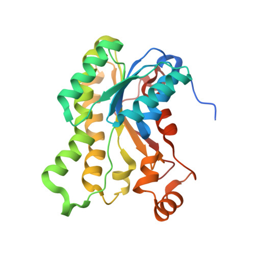
Crystal structure of L-arabinose 1-dehydrogenase as a short-chain reductase/dehydrogenase protein.

Watanabe, S.Yoshiwara, K.Matsubara, R.Watanabe, Y.

(2022) Biochem Biophys Res Commun 604: 14-21

  • DOI: https://doi.org/10.1016/j.bbrc.2022.03.028
  • Primary Citation Related Structures: 
    7WWX

  • PubMed Abstract: 

    l-Arabinose 1-dehydrogenase (AraDH) catalyzes the NAD(P) + -dependent oxidation of l-arabinose to L-arabinono-1,4-lactone in the non-phosphorylative l-arabinose pathway, and is classified into glucose-fructose oxidoreductase and short-chain dehydrogenase/reductase (SDR). We herein report the crystal structure of a SDR-type AraDH (from Herbaspirillum huttiense) for the first time. The interactions between Asp49 and the 2'- and 3'-hydroxyl groups of NAD + were consistent with strict specificity for NAD + . In a binding model for the substrate, Ser155 and Tyr168, highly conserved in the SDR superfamily, interacted with the C1 and/or C2 hydroxyl(s) of l-arabinose, whereas interactions between Asp107, Arg109, and Gln206 and the C2 and/or C3 hydroxyl(s) were unique to AraDH. Trp200 significantly contributed to the selectivities of the C4 hydroxyl and C6 methyl of substrates.


  • Organizational Affiliation
    • Faculty of Agriculture, Ehime University, 3-5-7 Tarumi, Matsuyama, Ehime, 790-8566, Japan; Department of Bioscience, Graduate School of Agriculture, Ehime University, 3-5-7 Tarumi, Matsuyama, Ehime, 790-8566, Japan; Center for Marine Environmental Studies (CMES), Ehime University, 2-5 Bunkyo-cho, Matsuyama, Ehime, 790-8577, Japan. Electronic address: irab@agr.ehime-u.ac.jp.

Macromolecule Content 

  • Total Structure Weight: 60.87 kDa 
  • Atom Count: 4,511 
  • Modeled Residue Count: 508 
  • Deposited Residue Count: 544 
  • Unique protein chains: 1

Macromolecules

Find similar proteins by:|  3D Structure
Entity ID: 1
MoleculeChains  Sequence LengthOrganismDetailsImage
NAD(P)-dependent dehydrogenase (Short-subunit alcohol dehydrogenase family)
A, B
272Herbaspirillum huttiense subsp. putei IAM 15032Mutation(s): 0 
Gene Names: DFS02_4196

Experimental Data & Validation

Experimental Data

  • Method: X-RAY DIFFRACTION
  • Resolution: 1.36 Å
  • R-Value Free:  0.192 (Depositor), 0.192 (DCC) 
  • R-Value Work:  0.168 (Depositor), 0.168 (DCC) 
  • R-Value Observed: 0.169 (Depositor) 
Space Group: C 1 2 1
Unit Cell:
Length ( Å )Angle ( ˚ )
a = 86.509α = 90
b = 83.562β = 113.04
c = 81.927γ = 90
Software Package:
Software NamePurpose
REFMACrefinement
XDSdata scaling
PDB_EXTRACTdata extraction
BALBESphasing

Structure Validation

View Full Validation Report


Ligand Structure Quality Assessment 


Entry History 

& Funding Information

Deposition Data


Funding OrganizationLocationGrant Number
Not funded--

Revision History  (Full details and data files)

  • Version 1.0: 2022-03-30
    Type: Initial release
  • Version 1.1: 2023-11-29
    Changes: Data collection, Refinement description