7WSG | pdb_00007wsg

Cryo-EM structure of SARS-CoV spike receptor-binding domain in complex with sea lion ACE2


Experimental Data Snapshot

  • Method: ELECTRON MICROSCOPY
  • Resolution: 3.03 Å
  • Aggregation State: PARTICLE 
  • Reconstruction Method: SINGLE PARTICLE 

Starting Model: experimental
View more details

wwPDB Validation 3D Report Full Report

Validation slider image for 7WSG

This is version 1.2 of the entry. See complete history

Literature

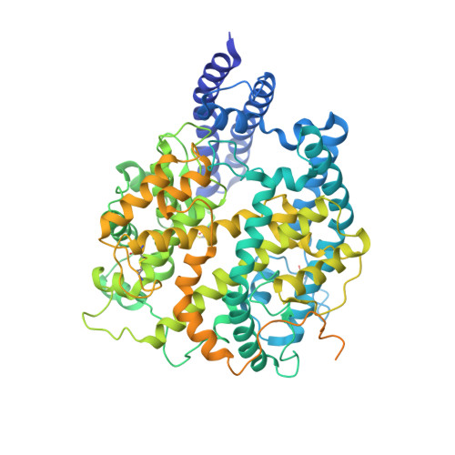
Cross-species recognition and molecular basis of SARS-CoV-2 and SARS-CoV binding to ACE2s of marine animals.

Li, S.Yang, R.Zhang, D.Han, P.Xu, Z.Chen, Q.Zhao, R.Zhao, X.Qu, X.Zheng, A.Wang, L.Li, L.Hu, Y.Zhang, R.Su, C.Niu, S.Zhang, Y.Qi, J.Liu, K.Wang, Q.Gao, G.F.

(2022) Natl Sci Rev 9: nwac122-nwac122

  • DOI: https://doi.org/10.1093/nsr/nwac122
  • Primary Citation Related Structures: 
    7WSE, 7WSF, 7WSG, 7WSH

  • PubMed Abstract: 

    Severe acute respiratory syndrome coronavirus 2 (SARS-CoV-2) has an extremely broad host range that includes hippopotami, which are phylogenetically closely related to whales. The cellular ACE2 receptor is one of the key determinants of the host range. Here, we found that ACE2s from several marine mammals and hippopotami could efficiently bind to the receptor-binding domain (RBD) of both SARS-CoV and SARS-CoV-2 and facilitate the transduction of SARS-CoV and SARS-CoV-2 pseudoviruses into ACE2-expressing cells. We further resolved the cryo-electron microscopy complex structures of the minke whale ACE2 and sea lion ACE2, respectively, bound to the RBDs, revealing that they have similar binding modes to human ACE2 when it comes to the SARS-CoV-2 RBD and SARS-CoV RBD. Our results indicate that marine mammals could potentially be new victims or virus carriers of SARS-CoV-2, which deserves further careful investigation and study. It will provide an early warning for the prospective monitoring of marine mammals.


  • Organizational Affiliation
    • CAS Key Laboratory of Pathogenic Microbiology and Immunology, Institute of Microbiology, Chinese Academy of Sciences, Beijing100101, China.

Macromolecule Content 

  • Total Structure Weight: 114.7 kDa 
  • Atom Count: 6,413 
  • Modeled Residue Count: 787 
  • Deposited Residue Count: 996 
  • Unique protein chains: 2

Macromolecules

Find similar proteins by:|  3D Structure
Entity ID: 1
MoleculeChains  Sequence LengthOrganismDetailsImage
Angiotensin-converting enzyme806MammaliaMutation(s): 0 
Gene Names: ACE2
EC: 3.4
UniProt
Find proteins for A0A6J2EID0 (Zalophus californianus)
Explore A0A6J2EID0 
Go to UniProtKB:  A0A6J2EID0
Entity Groups
Sequence Clusters30% Identity50% Identity70% Identity90% Identity95% Identity100% Identity
UniProt GroupA0A6J2EID0
Sequence Annotations
Expand
Reference Sequence
Find similar proteins by:|  3D Structure
Entity ID: 2
MoleculeChains  Sequence LengthOrganismDetailsImage
Spike protein S1190Severe acute respiratory syndrome coronavirusMutation(s): 0 
Gene Names: S2
UniProt
Find proteins for P59594 (Severe acute respiratory syndrome coronavirus)
Explore P59594 
Go to UniProtKB:  P59594
Entity Groups
Sequence Clusters30% Identity50% Identity70% Identity90% Identity95% Identity100% Identity
UniProt GroupP59594
Sequence Annotations
Expand
Reference Sequence

Small Molecules

Experimental Data & Validation

Experimental Data

  • Method: ELECTRON MICROSCOPY
  • Resolution: 3.03 Å
  • Aggregation State: PARTICLE 
  • Reconstruction Method: SINGLE PARTICLE 
EM Software:
TaskSoftware PackageVersion
MODEL REFINEMENTPHENIX

Structure Validation

View Full Validation Report


Entry History 

& Funding Information

Deposition Data


Funding OrganizationLocationGrant Number
Chinese Academy of SciencesChinaXDB29010202

Revision History  (Full details and data files)

  • Version 1.0: 2022-11-09
    Type: Initial release
  • Version 1.1: 2024-11-13
    Changes: Data collection, Refinement description, Structure summary
  • Version 1.2: 2025-07-02
    Changes: Data collection